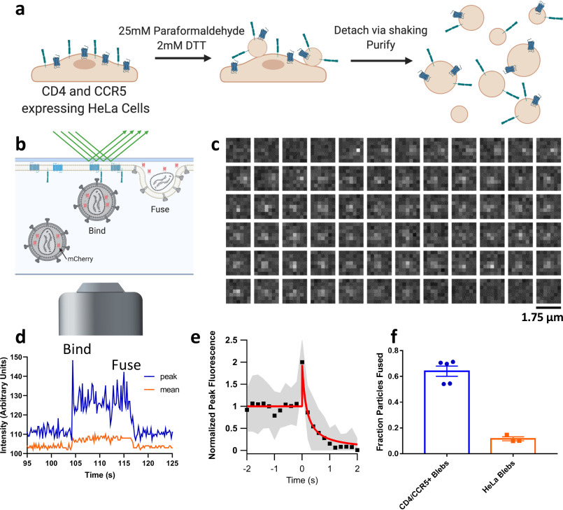
Figure 1.
Membrane blebs as a model for studying viral fusion. a, cartoon depicting the protocol for making CD4- and CCR5-containing blebs from HeLa cells. Detached blebs are mixed with HIV pseudoviruses and frozen for cryoET or used to form a SPPM as shown in b. b, cartoon showing discrete steps of HIV fusion to a bleb-derived SPPM in a TIRF-based single-particle fusion assay. All pseudoviruses are grown with a genetically encoded soluble content marker, mCherry, that upon fusion diffuses out of the virus and into the cleft between the SPPM and the substrate (described in Ref. 21). c, example micrographs of an HIV pseudovirus particle with a diffusible mCherry content marker fusing with a CD4 and CCR5 containing SPPM. Each box represents the same region separated in time by 0.2 s. d, fluorescence intensity of the same particle is plotted over time, where peak is the intensity of the brightest pixel in the 7 × 7 region and mean is the average intensity of the same area. e, 30 intensity traces of fusing particles were aligned to the increase in intensity at the onset of fusion, averaged (black squares show the mean, gray shaded area shows S.D.), and fit to a release model as shown in Fig. S1 (red line). f, fraction of stably bound particles that undergo fusion to SPPMs made with blebs from CD4- and CCR5-overexpressing HeLa cells or HeLa cells that do not express either. Each point represents a separately prepared bilayer. Error bars, S.E.

