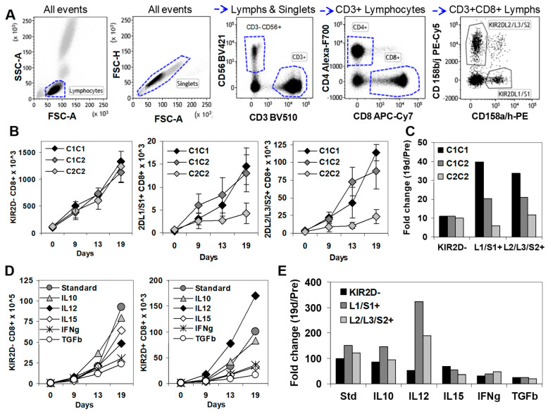
Figure 4.
In vitro expansion of KIR2D− and KIR2D+ CD8+ T cells. (A) Immunophenotype analysis of KIR2D− and KIR2D+ CD8+T cells. Left to right, FSC/SSC dot plot showing all events to select lymphocytes, FSC-A/FSC-H dot plot showing all events to select singlets, CD3/CD56 dot plot showing singlet/lymphocytes to select CD3+ T cells and CD3−CD56+ NK cells, CD4/CD8 dot plot showing CD3+ lymphocytes to select CD4+ and CD8+ T cell subsets, and CD158ah/CD158bj dot plot showing CD8+ lymphocytes to select CD158ah (KIR2DL1/S1+) and CD158bj (KIR2DL2/L3/S2+) CD8+ T cells. (B) In vitro expansion of KIR2D− and KIR2D+ CD8+ T cell subsets from peripheral blood mononuclear cells (PBMC) of C1C1, C1C2, and C2C2 healthy donors in the presence of IL-2 (40 µg/mL), 0.5 × 10⁶ 100 Gy irradiated JY cell line, and 5 × 10⁶ 25 Gy irradiated PBMC from the same C1C2 healthy donor, at days 0, 9, 13, and 19 (standard conditions). These experiments were repeated 3 times. (C) Fold increase at day 19 of CD8+ T cell subsets in C1C1, C1C2, and C2C2 donors. (D) In vitro expansion of KIR2DL1/S1+ and KIR2DL2/L3/S2+ CD8+T cell subsets from PBMC of a C1C2 donor in the same conditions as in B, but IL-10 (20 ng/mL), IL-12 (5 ng/mL), IL-15 (10 ng/mL), IFNγ (50 ng/mL), or TGFβ (1 ng/mL) were added at day 0. (E) Fold change at day 19 of CD8+ T cell subsets after the addition of different cytokines to the standard expansion protocol.

