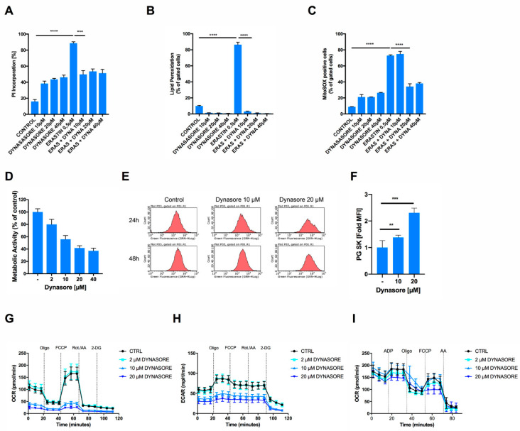
Figure 3.
Dynasore protects neuronal cells from ferroptosis and inhibits mitochondrial respiration. (A,B) HT22 cells were treated with DMSO and with the indicated concentrations of dynasore and erastin (0.5 µM) for 16 h. Cell death was determined by AnnexinV/PI staining and fluorescence-activated cell sorting (FACS) measurements. Lipid peroxidation was quantified by BODIPY C11 staining and flow cytometry after 8 h of treatment. (C) HT22 cells were treated with DMSO, the indicated concentrations of dynasore and erastin (0.5 µM) for 16 h. MitoSOX-positive cells were gated and quantified by flow cytometry. (D) HT22 cells were incubated with the indicated concentrations of dynasore for 16 h. Metabolic activity was determined by MTT assay. (E,F) HT22 cells were treated as indicated for 24 h, stained by Phen Green SK staining (PG SK) (5 µM) and fold mean fluorescence intensity (MFI) was recorded by flow cytometry. (G,H) HT22 cells were treated with dynasore and erastin for 16 h. Measurement of the oxygen consumption rate (OCR) and extracellular acidification rate (ECAR) were determined simultaneously by seahorse assay. After recording of three baseline measurements, injections were performed as follows: (Oligo) 3 µM oligomycin, (FCCP) 0.5 µM FCCP, (Rot./AA) 100 nM Rotenone/1 µM Antimycin A, (2-DG) 50 mM 2-DG. (I) Sprague Dawley rat mitochondria were isolated, treated with dynasore for 1 h at the indicated concentrations and OCR was recorded by Seahorse Assay as in G. All data are means +/− SD or representative images where applicable. * indicates p < 0.05; ** indicates p < 0.01; *** indicates p < 0.001; **** indicates p < 0.0001; ns indicates non-significant differences.

