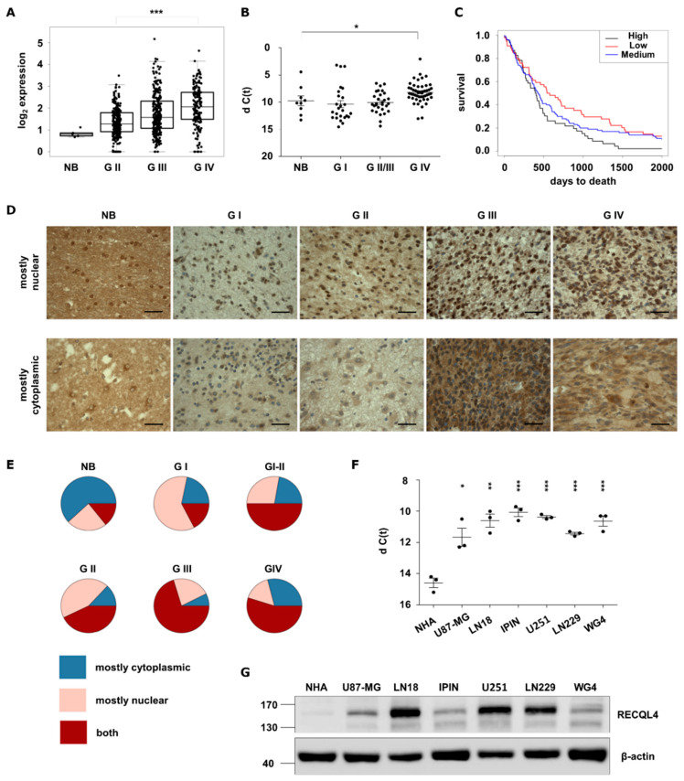
Figure 1.
RECQL4 expression is upregulated in human malignant gliomas. (A) RECQL4 expression in normal brain (NB), WHO grade II and grade III gliomas and glioblastomas (GBM, WHO grade IV) in TCGA datasets. Presented values are log2 of FPKM values. Statistical significance was determined by Welch’s analysis of variance (ANOVA) between GII, GIII and GIV groups. (B) Quantitative analysis of RECQL4 mRNA levels in NB (n = 9), and gliomas of different grades: GI (n = 25), GII/III (n = 29) and GBM (n = 50). The RECQL4 expression was normalized to GAPDH; results represent means ± SEM; statistical significance was determined by one-way ANOVA, followed by Dunnett’s post hoc test. p-Values were considered as significant when * p < 0.05. (C) Kaplan–Meier overall survival analysis of LGG and GBM patients from TCGA. Log-rank test was calculated between RECQL4 LOW and HIGH expression groups (* p < 0.05). (D) Representative immunostaining showing expression of RECQL4 protein in the glioma tissue microarray including astrocytomas (n = 132), glioblastomas (n = 31), oligoastrocytomas (n = 7), oligodendrogliomas (n = 9), ependymomas (n = 11), ganglioglioma (n = 1) and gliosarcoma (n = 1), plus tumour adjacent and normal brain (NB) tissues (n = 8). RECQL4 expression appeared as high nuclear and low cytoplasmic signal (upper panel) and low nuclear and high cytoplasmic signal (bottom panel). (E) Quantification of RECQL4 immunoreactivity. Statistical significance was determined by chi-squared test. p-values of NB p = 1.0, GI p = 0.019, GII p = 1.1 × 10⁻⁵, GIII p = 6.3 × 10⁻⁵, GIV p = 0.011. (F) Quantitative analysis of RECQL4 mRNA levels in established glioma cell lines, patient-derived primary cultures, and normal human astrocytes (NHA). The expression was normalized to GAPDH; results represent means ± SEM of 3 cell passages (n =3). Statistical significance was determined by one-way ANOVA. p-Values were considered as significant when * p < 0.05, ** p < 0.01, *** p < 0.001. (G) Representative immunoblot shows RECQL4 expression in established glioma cell lines and GBM primary cultures in comparison to NHA. β-Actin was used as a loading control.

