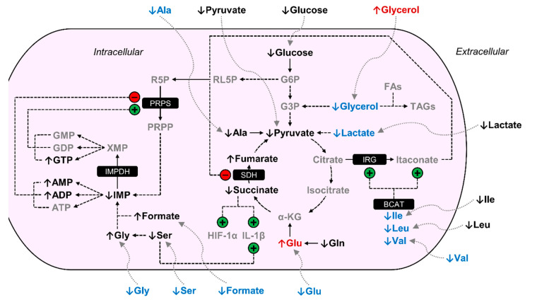
Figure 3.
BCM-exposed MΦs are metabolically distinct from PCM-exposed MΦs. Arrows adjacent to metabolite names are indicative of the metabolite level in BCM-exposed MΦs relative to PCM-exposed MΦs. Metabolites in gray were not detected in our NMR spectra. Metabolites in black were detected but were not significantly increased or decreased in BCM-exposed MΦs relative to PCM-exposed MΦs. Metabolites in red were significantly elevated in BCM-exposed MΦs relative to PCM-exposed MΦs, and metabolites in blue were significantly reduced in BCM-exposed MΦs relative to PCM-exposed MΦs. Regulation demonstrated by previous studies that are mentioned in the discussion are indicated with circles containing either a +/green for stimulation or -/red for inhibition. Abbreviations denote: ADP, adenosine diphosphate; Ala, alanine; AMP, adenosine monophosphate; ATP, adenosine triphosphate; BCAT, branched-chain aminotransferase; FAs, fatty acids; G3P, glyceraldehyde 3-phosphate; G6P, glucose 6-phosphate; GDP, guanosine diphosphate; Gln, glutamine; Glu, glutamate; Gly, glycine; GMP, guanosine monophosphate; GTP, guanosine triphosphate; HIF-1α, hypoxia-inducible factor-1α; IL-1β, interleukin-1β; IMP, inosine monophosphate; IMPDH, IMP dehydrogenase; IRG, immune-responsive gene; Ile, isoleucine; Leu, leucine; PRPP, 5-phosphoribosyl-1-pyrophosphate; PRPS, PRPP synthetase; R5P, ribose 5-phosphate; RL5P, ribulose 5-phosphate; SDH, succinate dehydrogenase; Ser, serine; TAGs, triacylglycerides; Val, valine; XMP, xanthosine monophosphate; α-KG, α-ketoglutarate.

