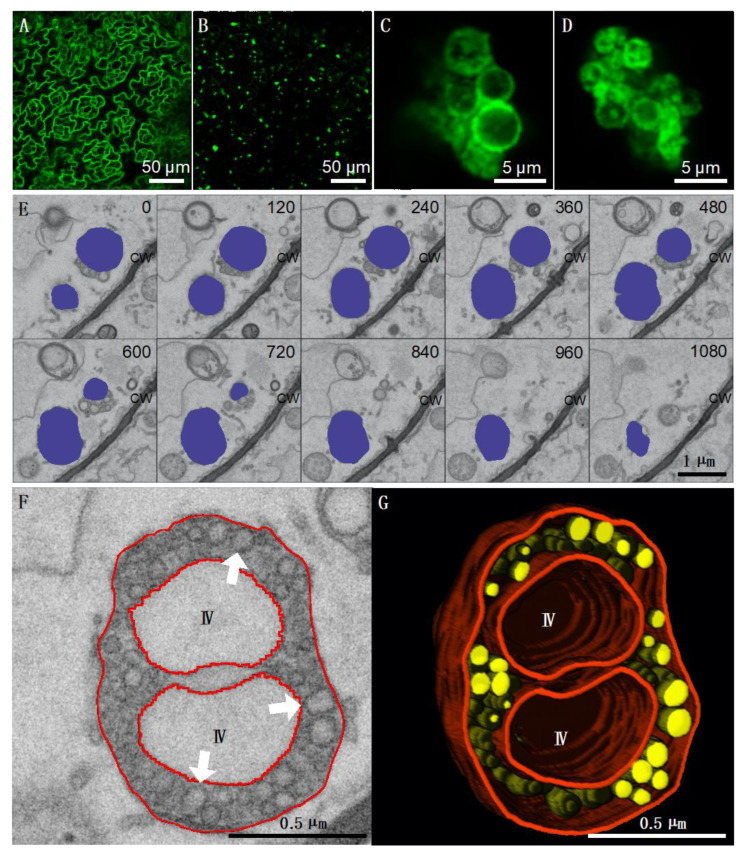
Figure 2.
TBSV replicates in association with peroxisomes in Arabidopsis thaliana. Confocal microscopy imaging of leaf epidermal cells from healthy (A) and TBSV-infected (B–D) 35S:B2:GFP A. thaliana Col-0 plants at 9 dpi. At low magnification (A,B), B2:GFP adopts a nucleo-cytoplasmic localization in healthy cells (A), whereas in TBSV-infected tissues, B2:GFP is relocalized to numerous cytoplasmic aggregates (B). At high magnification (C,D), B2:GFP is essentially found in association with clustered ring-like structures. (E) Array of individual serial block face SEM (SBEM) images taken from 35S:B2:GFP A. thaliana plants infected with TBSV at 9 dpi. Image planes were recorded at 30 nm intervals, and the entire acquisition was recorded using 150 image planes. Ten sample slices taken at 120 nm intervals are shown in (E). Altered peroxisomes containing numerous spherules are shaded in blue. (F) Detail of an altered peroxisome in which numerous spherules are visible (white arrows). (G) Partial 3D reconstruction of the same peroxisome in which spherules are represented in yellow and membranes in red. IV: Intraperoxisomal vacuoles. CW: Cell wall.

