Figure 7.
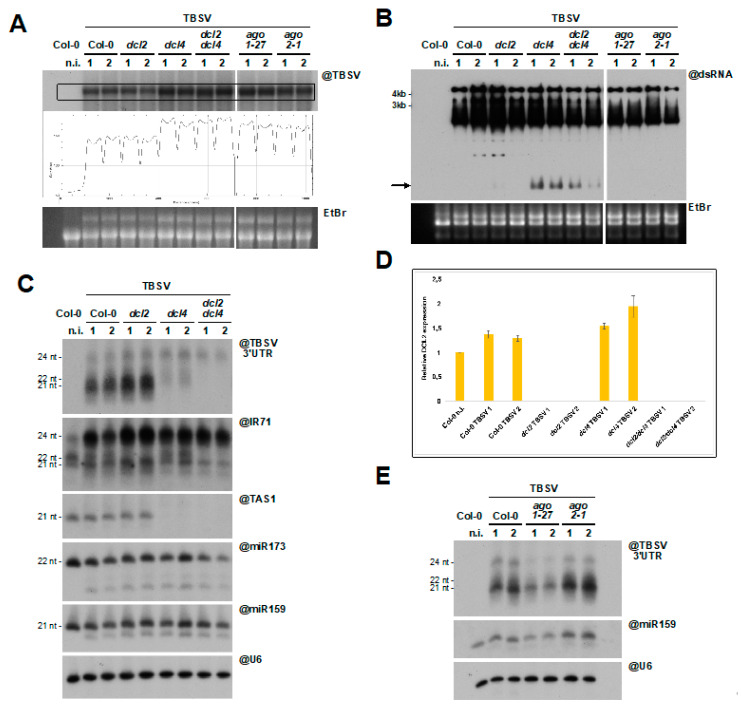
Atypical RNA silencing response to TBSV infection in A. thaliana. (A) Northern blot of high molecular weight RNA to detect TBSV in systemically infected rosette leaves, 10 dpi, of A. thaliana mutant lines. Quantification of signal intensity is provided. Each sample is a pool of 4–5 plants. (B) Northwestern blot to detect dsRNA in the samples described in (A). The black arrow indicates the dsRNA band observed upon genetic knock-out of DCL4. In (A,B) EtBr gel staining was used as a loading control. (C) Northern blot analysis of low molecular weight RNA to detect TBSV-derived and endogenous small RNA. The same membrane was repeatedly stripped and re-probed. (D) RT-qPCR of the samples analyzed in (A–C) to assess accumulation of DCL2 mRNA. Expression levels were normalized to UBIQUITIN 10 (UBQ10) and ACTINE2 mRNA. Primers were designed on the opposite sides of the dcl2-1 T-DNA insertion to rule out non-specific amplification. (E) Northern blot analysis of low molecular weight RNA to detect TBSV-derived siRNA in the ago mutants analyzed in (A,B). As in (C), miR159 and snU6 were used as loading controls.

