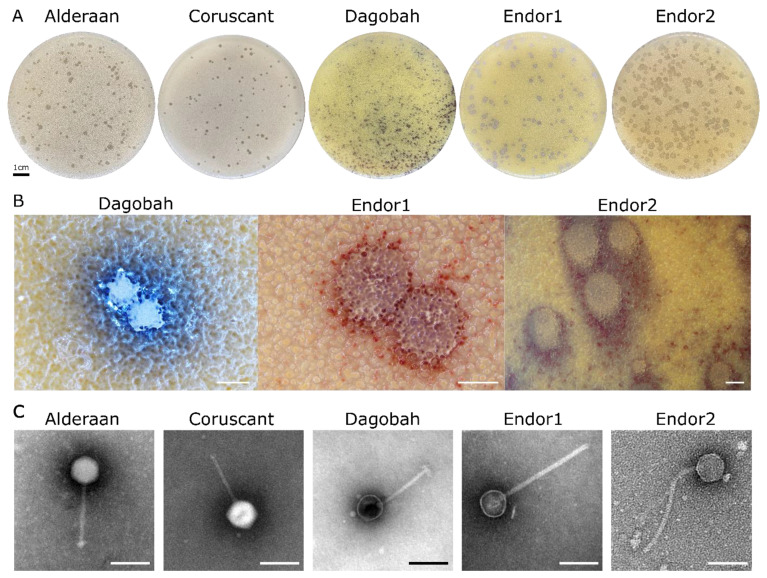
Figure 1.
Morphology observation of five novel Streptomyces phages. (A) Plaque morphologies of the five phages. Double agar overlays were performed to infect S.venezuelae ATCC 10712 with the phages Alderaan and Coruscant, and S. coelicolor M600 with the phages Dagobah, Endor1 and Endor2. Plates were incubated overnight at 30 °C and another day (3 days in the case of Dagobah) at room temperature to reach full maturity of the bacterial lawn; (B) Close-ups of phage plaques imaged using a stereomicroscope Nikon SMZ18. S. coelicolor M145 was infected by phages using GYM double agar overlays. The plates were incubated at 30 °C overnight and then kept at room temperature for two (Endor1 and Endor2) or three days (Dagobah). Scale bar: 1 mm; (C) Transmission electron microscopy (TEM) of phage isolates. The phage virions were stained with uranyl acetate. Scale bar: 150 nm.

