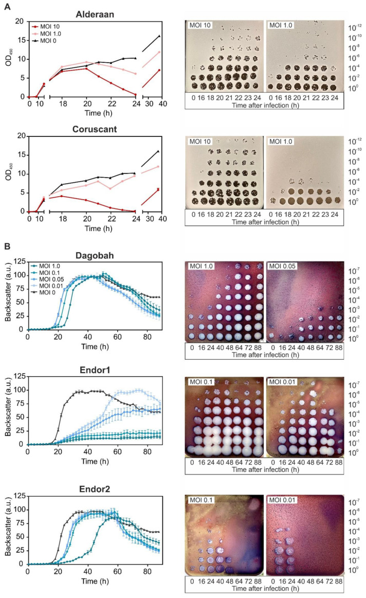
Figure 2.
Infection curves of the five phages infecting S. venezuelae (A) and S. coelicolor (B). Spores of either S. venezuelae (105) or S. coelicolor M145 (106) were grown in GYM or YEME medium, respectively. After 6 to 8 h, phages were added at the corresponding multiplicity of infection (MOI). OD450 or backscatter were measured over time (left panels), in parallel to phage titers (right panels).

