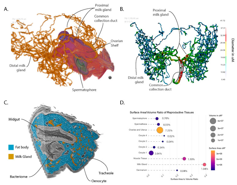
Figure 5.
Views of milk gland morphology and structural features with other reproductive and abdominal features for context. (A) Orthogonal view with sagittal section through uterus, ovaries and spermatheca; (B) Dorsal view of a thickness mesh of the milk gland; (C) Orthogonal view of the abdominal volume with milk gland and fat body denoted by color; (D) Quantification and visualization of relative volumes, surface areas, surface area to volume ratios and percentage to total abdominal volume within the scan. Circle sizes represent volume in µm3, circle color represents surface area in µm2, percentages represent proportion of the total volume of the abdominal tissue included within the scan.

