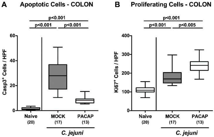
Figure 4.
Apoptotic and proliferating colonic epithelial cells following PACAP treatment of C. jejuni infected secondary abiotic IL-10−/− mice. Mice were perorally infected with C. jejuni strain 81–176 by gavage on day (d) 0 and d1, and subjected to intraperitoneal treatment, with either synthetic PACAP or vehicle (mock) from d2 until d5 post-infection. On d6, the average numbers of (A) apoptotic (cleaved caspase-3 positive, Casp3+) and (B) proliferating (Ki67+) epithelial cells were quantitated in six high power fields (HPF) of colonic paraffin sections applying immunohistochemistry. Naive mice were used as negative controls. Box plots represent the 75th and 25th percentiles of the median (black bar inside the boxes). The total range, the significance levels (p values calculated by the one-way ANOVA test followed by the Tukey post-correction test for multiple comparisons) and the total numbers of mice under investigation (in parentheses) are indicated. Results pooled from four independent experiments are shown.

