Figure 1.
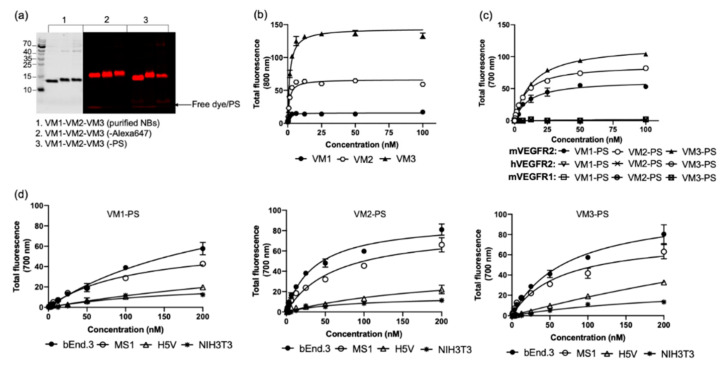
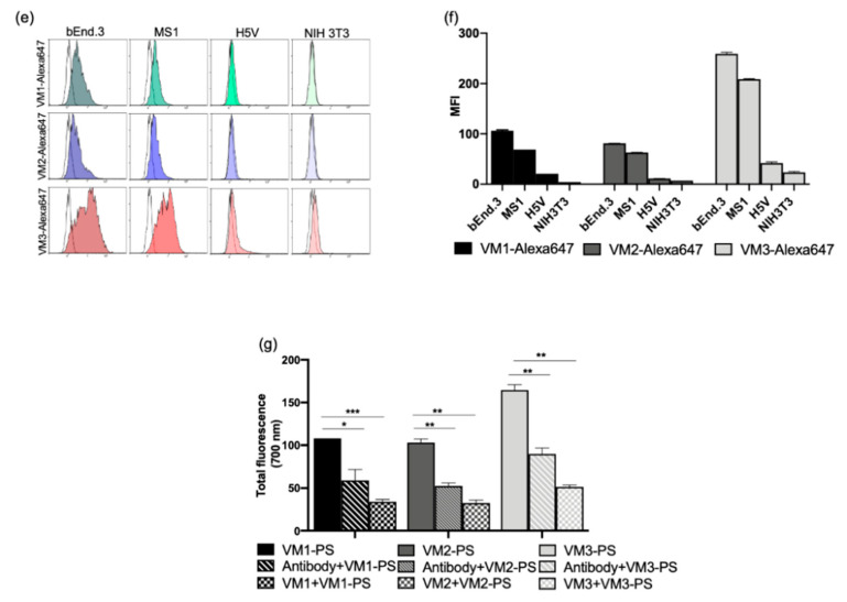
Purity and specificity of the NBs and NB–conjugates. (a) Purified NBs, NB–Alexa647, and NB–PS conjugates separated by SDS-PAGE. Free dye/PS is observed at the gel front (arrow): (1) purified NBs after PageBlue staining (depicted in black); and (2,3) the fluorescence of Alexa647 and PS detected at 700 nm, respectively (depicted in red). (b) Binding of the unlabeled NBs to the mVEGFR2 protein detected by anti-VHH antibody. (c) Binding of the NB–PS conjugates to the mVEGFR2, hVEGFR2, and mVEGFR1 proteins. Total fluorescence of NB–PS bound to the protein was detected using an Odyssey infrared scanner at 700 nm. (d) Total fluorescence intensity of cell bound/internalized NB–PS conjugates on the murine cell lines after 1-h incubation at 37 °C. (e,f) Fluorescence of NB–Alexa647 conjugates detected by flow cytometry. The murine cell lines were incubated with the conjugates for 1 h at 37 °C followed by trypsinization and FACS analysis. Mean fluorescent intensity (MFI) obtained from flow cytometry. Data are shown as mean ± SD. (g) In vitro competition experiments of the NB–PS conjugates tested on bEnd.3 cells in the presence or absence of ten-times excess of anti-VEGFR2 antibody or unconjugated NBs. Total fluorescence intensity of the associated NB–PS conjugates was detected by an Odyssey infrared scanner at 700 nm (* p < 0.05; ** p < 0.01; *** p < 0.001; t-test).


