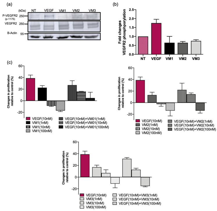
Figure 3.
Anti-VEGFR2 NBs blocked VEGF-induced proliferation and did not act as receptor agonists. (a) VEGF-A (50 nM) or NBs (1 µM) were added to the serum-starved MS1 cells and incubated for 15 min. VEGFR2 phosphorylation was measured in the total cell lysates by Western blotting: (top) staining of phosphorylated tyrosine 1175 of VEGFR2; (middle) staining of total VEGFR2; and (bottom) staining of actin. (b) Fold changes of P-VEGFR2 in MS1 cells treated with VEGF or nanobodies relative to the non-treated cells (NT). (c) MS1 cells treated with VEGF-A/NBs or both for 72 h followed by viability assay using AlamarBlue® reagent. Data are presented as percent changes in cell proliferation relative to the non-treated cells (mean ± SD).

