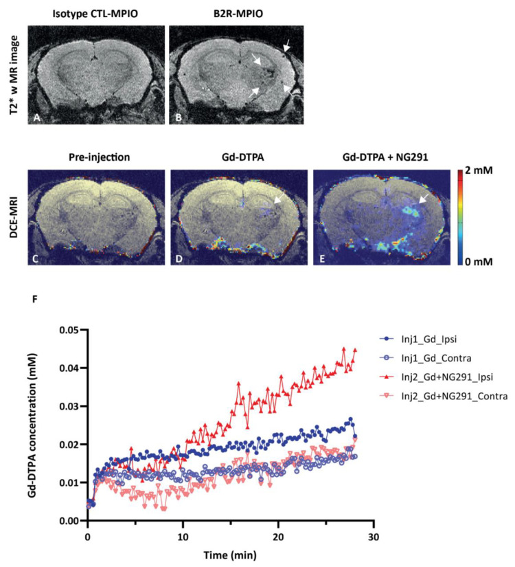
Figure 5.
BBB modulatory effects of the B2R agonist analog NG291 on irradiated mouse brain. (A,B) Representative in vivo T2*-weighted 3D MR coronal image after (A) isotype control-microparticles of iron oxide (MPIO) or (B) B2R-MPIOs-injection. At 10 months after irradiation, specific retention of the contrast agent on activated vasculature is visible as signal voids (B, white arrow) in the right hemisphere, with little negative contrast observed in the contralateral control hemisphere or (A) with CTL-MPIO injection. (C–E) MRI assessment of changes in mouse brain vascular permeability after irradiation. Representative in vivo T1-weighted color-coded coronal images (C) before and (D) after Gd-DTPA injection and (E) after NG291 and Gd-DTPA. White arrows in D and E indicate the location of increased vascular permeability to Gd-DTPA. (F) Magnevist (Gd-DTPA) uptake in the irradiated (ipsilateral) and nonirradiated (contralateral) hemisphere as a function of time, before and after infusion of NG291. Signal increase in the irradiated hemisphere is at least 2-fold higher after B2R agonist injection. Pseudo-color bar indicates Gd-DTPA concentration.

