Figure 2.
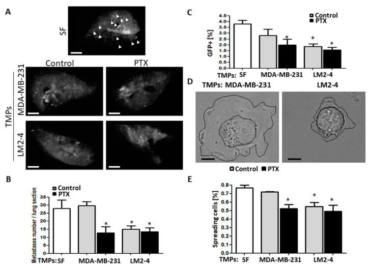
TMPs from highly metastatic cells or cells exposed to chemotherapy decrease tumor cell seeding. (A–C) GFP-positive MDA-MB-231 cells cultured with serum-free medium or TMPs from MDA-MB-231 or LM2-4 cells exposed to PTX or vehicle control were assessed for their seeding properties using the ex vivo pulmonary metastasis assay (PuMA), as described in Materials and Methods. Lung sections (n = 4 mice/group) were imaged (A) and the number of metastatic foci were counted (B). Scale bar, 0.2 cm. White arrows point at metastatic foci only in the serum-free group. The percentage of metastatic cells in the lungs was also assessed by flow cytometry after the lung sections underwent single cell suspension (C). (D,E) Cells (treated as in A–C) were seeded on fibronectin-coated plates. Cell spreading was then immediately assessed by time-lapse microscopy, in which the area of cell spread was displayed. Representative images are shown in (D). Scale bar, 20 µm. Cell cytoplasmic membrane and nucleus are drawn in black lines. The percentage of spreading cells when cultured on fibronectin is shown in (E). n = 3 repeats/group with the assessment of ~100 cells/repeat. * differences compared to MDA-MB-231 control group, *, p < 0.05, as assessed by one-way ANOVA followed by Tukey post-hoc test.

