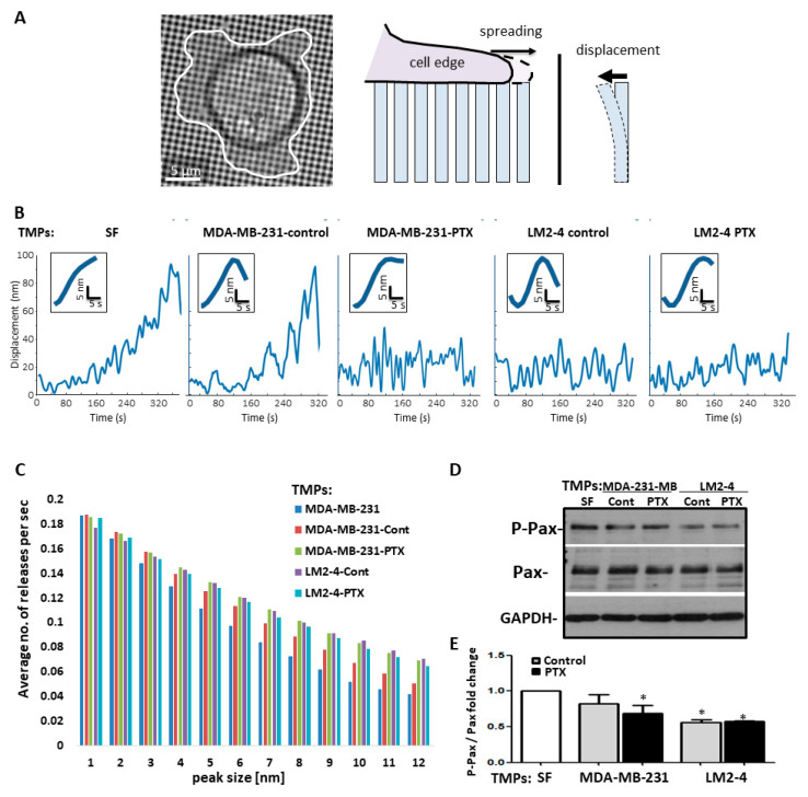
Figure 4.
TMPs from highly metastatic cells or cells exposed to chemotherapy increase the pace of cell biomechanical forces and inhibit focal adhesion signaling. (A–C) MDA-MB-231 cells cultured with TMPs from MDA-MB-231 or LM2-4 cells exposed to PTX or vehicle control were applied on a pillar assay, as described in Materials and Methods. MDA-MB-231 cells cultured with serum-free medium (SF) were used as a control. (A) Left: Example of a cell spreading on an array of 0.5 µm diameter fibronectin-coated pillars (see illustration on the right). (B) Typical pillar displacement curves. (C) Histograms presenting the average number of pillar releases per second binned according to the size of the release. Each of the control conditions was significantly different than the conditions in which the cells were exposed to TMPs from LM2-4 cells or from cells exposed to PTX (α < 0.01; Kolmogorov–Smirnov test). n = 8–10 repeats/group. (D–E) Cells treated as above were seeded on fibronectin-coated plates to evaluate phospho-paxillin (P-Pax) and total-paxillin (Pax) expression by Western blot. GAPDH was used as a loading control (D). The ratio between P-Pax over Pax was calculated based on densitometry, and presented as fold change, for three biological repeats (E). n = 3 repeats/group. * differences compared to MDA-MB-231 control group, * p < 0.05, as assessed by one-way ANOVA followed by Tukey post-hoc test.

