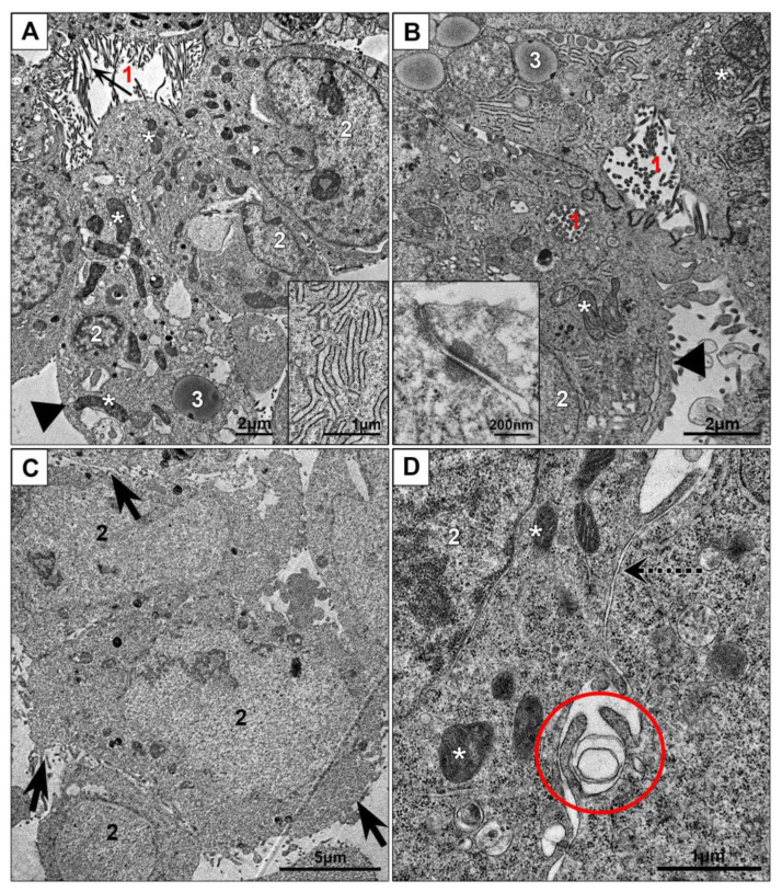
Figure 2.
Cell cultures in monolayer: (A,B)—HepG2, (C,D)—HEK293. Inserts: (A) cisternae of endoplasmic reticulum; (B) tight junction and desmosome between cells at apical pole. 1—“bile capillaries”, arrows show microvilli; 2—nucleus; 3—lipid droplet; asterisks show mitochondria; arrowheads show basolateral membranes; dotted arrow shows area of “simple” contact of two cells; oval shows site of macropinocytosis; tick arrows show cell surface with many outgrowths.

