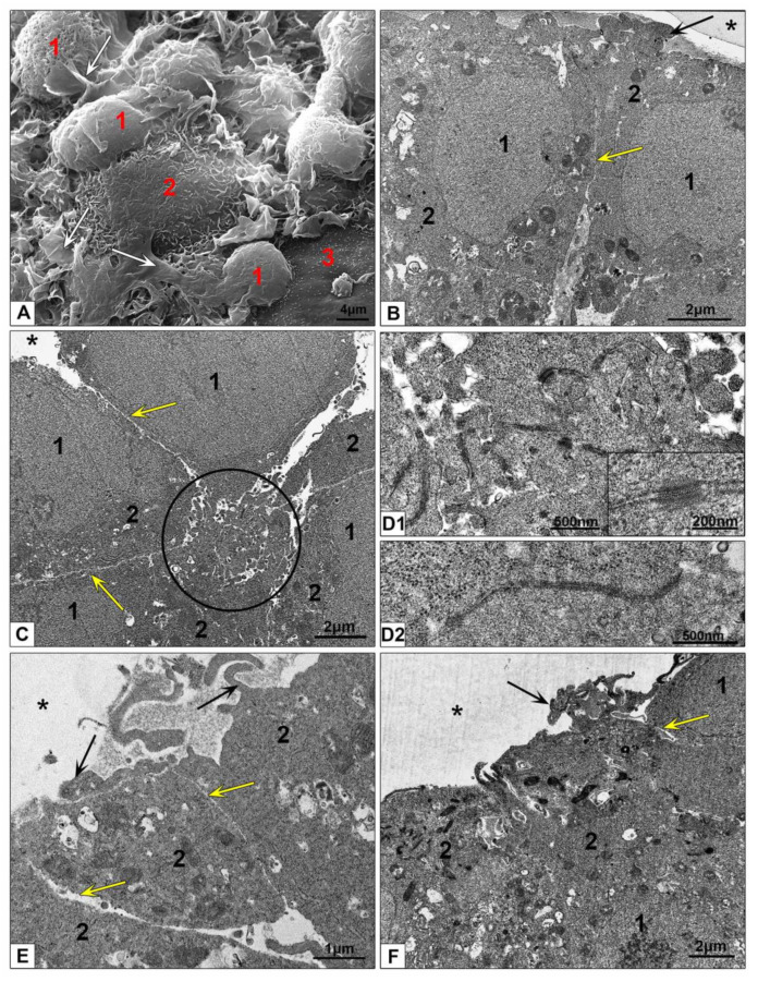
Figure 5.
Ultrastructure of HEK293 spheroids. (A) Representative SEM image of spheroid surface. 1—cell body; 2—cell surface with small microvilli; 3—flat cell surface; white arrows show flat folds. (B–F) Cells at the periphery of spheroid, ultrathin sections. 1—nucleus; 2—cytoplasm; asterisks show external space; circle shows a conglomerate of apical outgrowths; arrows show outgrowths protruding in external space; yellow arrows show narrow space between lateral cell surfaces. (D1) Outgrowth conglomerate at higher magnification, the insert shows desmosomes; (D2) this photo presents a structure similar to apical tight junction.

