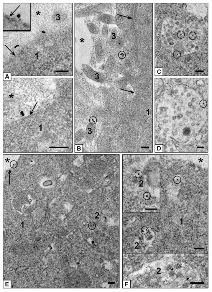
Figure 9.
Interaction of AuBSA-NPs with cells of different spheroids. (A–D) Penetration of AuBSA-NPs into HepG2 cells. (A,B) adsorption of NPs on plasmalemma, insert shows NP adsorption at high magnification. (C,D) AuBSA-NPs in late endosomes. (E,F) Penetration of AuBSA-NPs into HEK293 cells. (E) NPs on plasmalemma and in late endosome. (F) AuBSA-NPs in late endosomes and in a vesicle. 1—cytoplasm; 2—late endosomes; 3—cell outgrowths; asterisks show external space and arrows show plasmalemma. Bars correspond to 100 nm.

