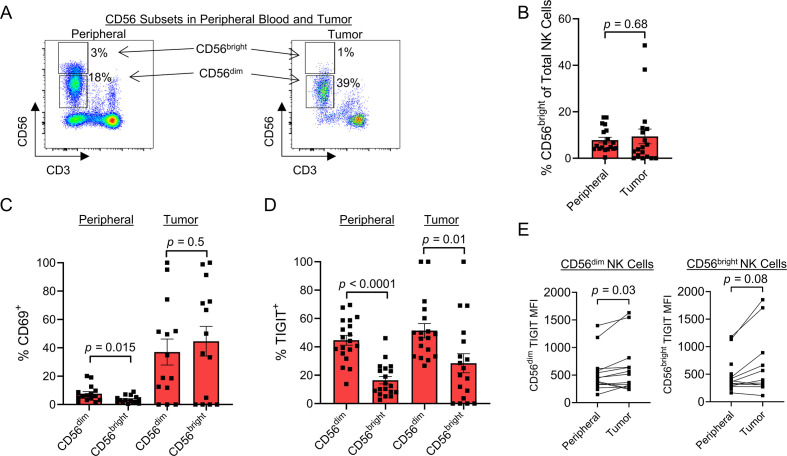
Figure 4.
CD56dim subset is the predominate NK cell within soft tissue sarcomas and expresses higher TIGIT. (A) Representative flow cytometry gating showing the CD56bright and CD56dim NK cell populations in the peripheral blood (left) and tumor (right). (B) No difference in proportion of NK cell subsets between the periphery and tumor. (C) Within the peripheral blood, CD56dim NK cells expressed higher levels of activation marker CD69 compared with CD56bright NK cells (p=0.015), which was not evident within the tumor. (D) Within the periphery (p<0.0001) and within the tumor (p=0.01), TIGIT expression was significantly higher on CD56dim NK cells compared with CD56bright NK cells. (E) CD56dim NK cell subset significantly increases TIGIT expression by MFI within the tumor (p=0.03). Mean±SEM. P values determined using paired Student’s t-test or Wilcoxon test. MFI, median fluorescence intensity; NK, natural killer.

