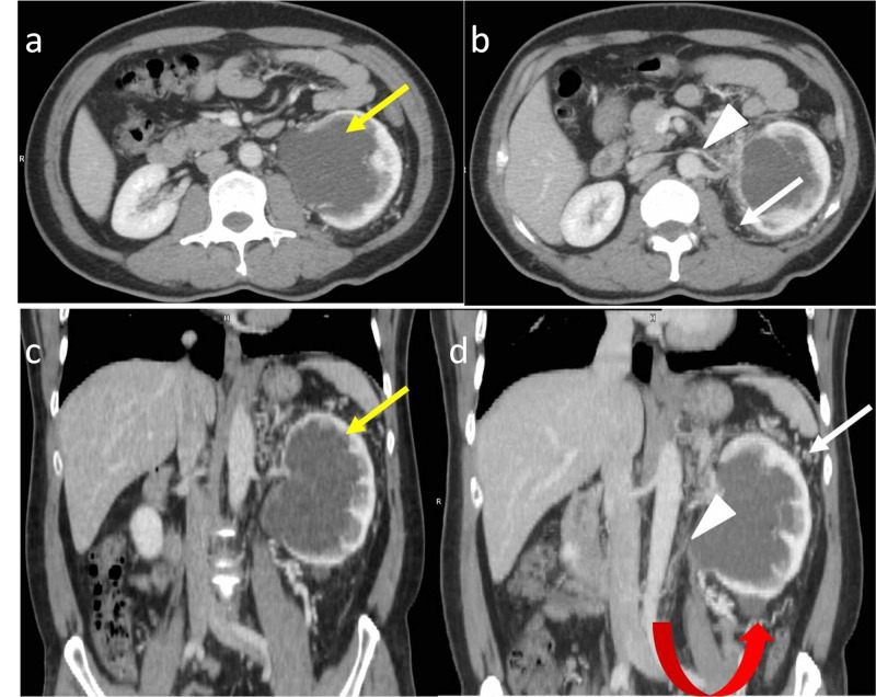
Figure 2. 49-year-old male with left peripelvic renal lymphangiectasia.
Contrast-enhanced CT of the abdomen in the venous phase in axial (a, b) and coronal planes (c, d) demonstrates a peripelvic cystic lesion (yellow arrows) on the left side. It also demonstrates the attenuated caliber of the left renal vein (white arrowheads) with perirenal collaterals (white arrows) consistent with chronic left renal vein thrombosis. Associated Bosniak type I cyst (red curved arrow) is seen in the lower pole of the left kidney
CT: computed tomography

