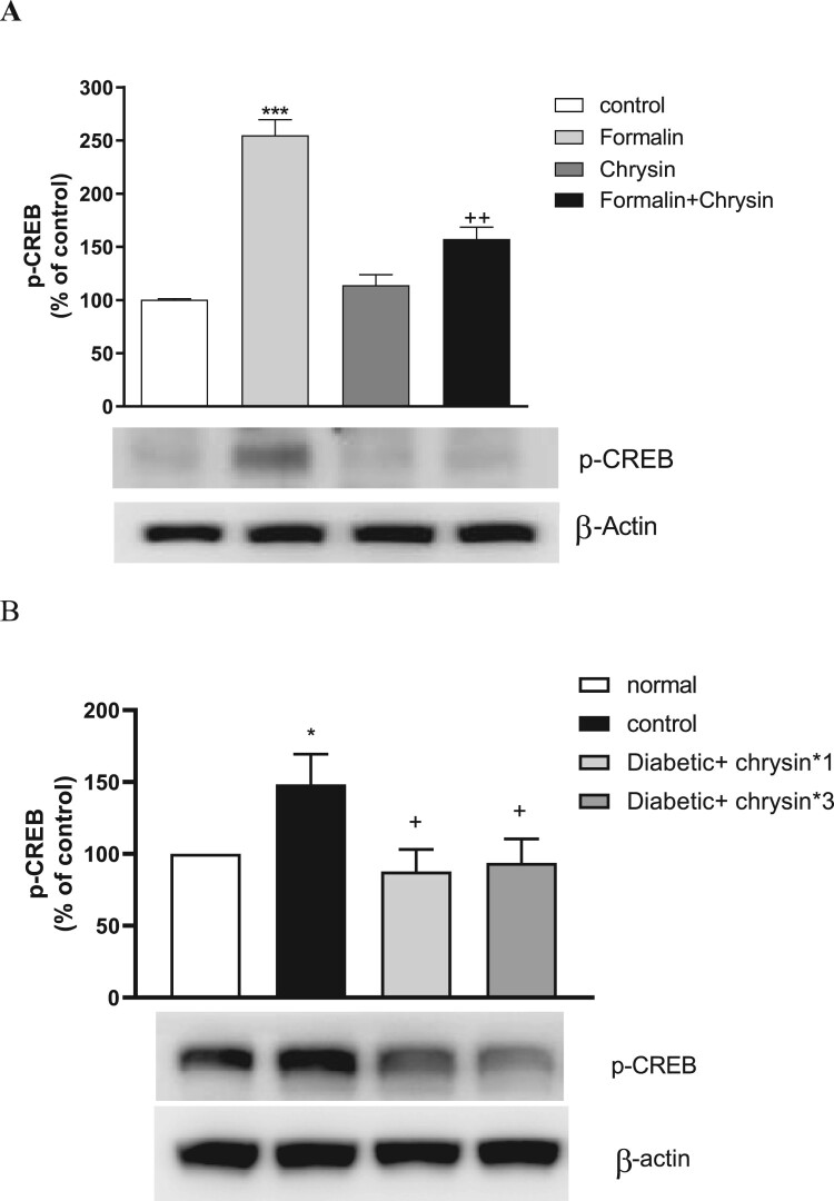
Figure 3.
Changes of phosphorylated CREB protein in the spinal cord by chrysin in the formalin-induced pain and diabete-induced nueropathy pain models. A. The proteins were extracted from dissected lumbar spinal cord 30 min after formalin injection for Western blot analysis. The number of animals in each group is 6. B. The experiment was performed at 5 weeks after streptozotocin injection. The diabetic mice were treated for chrysin for once or 3 times. The 3 times treatment of chrysin was performed twice each day (10 am and 4 pm). The proteins were extracted from dissected lumbar spinal cord 1hr after chrysin oral administration for Western blot analysis. β-Actin (1:1000 dilution) was used as an internal loading control. Signals were quantified with the use of laser scanning densitometry and expressed as a percentage of the control. Values are mean ± SEM (***P < 0.001, compared to Control group; ++P < 0.01, compared to formalin-treated group; *P < 0.05, compared to Normal group; +P < 0.05, compared to Diabetic group).

