
Figure 2.
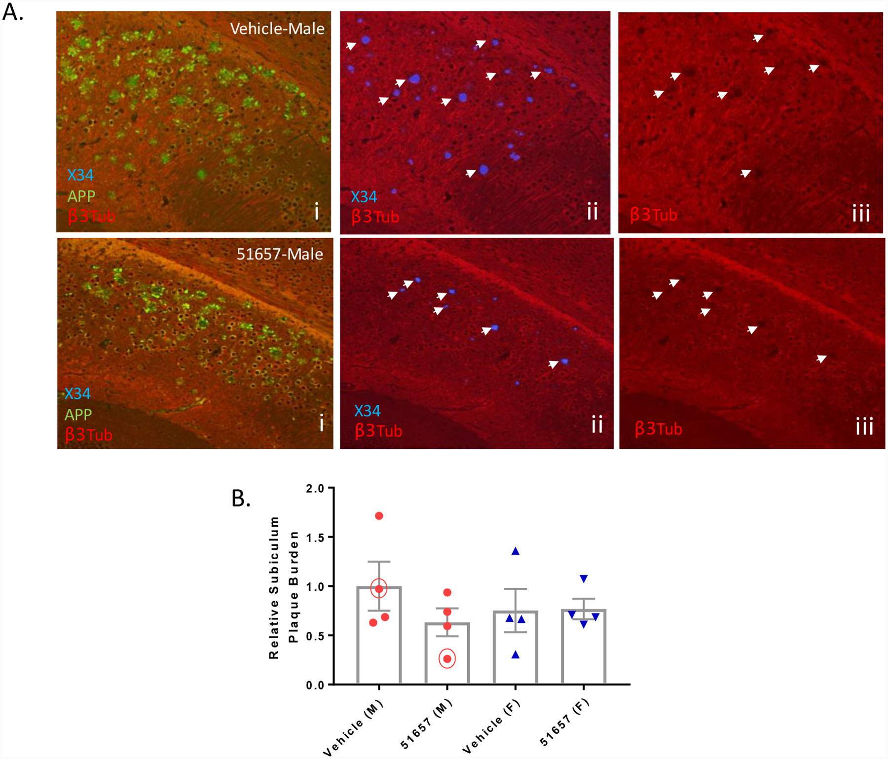
Male 5XFAD mice with decreased X34-positive SPs have decreased focal MT disruptions. A. Sections from the subiculum (bregma −3.08) of 5XFAD study mice i. stained for Aβ plaques (X34), APP-positive dystrophic processes and MTs (βIII-tubulin) show evidence of SP-associated voids of βIII-tubulin staining, indicative of MT disruption (plaques highlighted with arrowheads in panels ii with associated βIII-tubulin voids at identical sites shown in iii). The number of MT voids were reduced in male mice with reduced X34-positive plaques and APP-positive processes (compare upper and lower panels). B. Quantification of X34-positive plaques in the subiculum of the 5XFAD study mice after normalization to the mean of the male vehicle group, with n=4 vehicle- and 51657-treated male and female 5XFAD mice. The trend toward decreased plaque burden in the male 51657 treatment group did not reach statistical significance by unpaired 2-tailed t-test (t=1.28, df=6). Circled data points represent the mice shown in A. Error bars represent SEM.