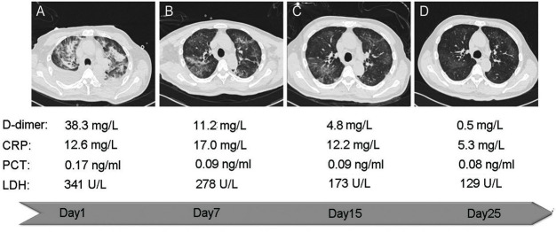
Fig 3. Representative computed tomography of a 62-year-old male patient during adjuvant antithrombotic treatment.
Abidol and Ibuprofen was taken orally by the patient intermittently. After admission, the combination of anticoagulation was used (Aspirin enteric-coated tablets 100mg once a day and Clopidogrel bisulfate tablet 75mg once a day). The representative CT and the blood test results during the treatment were shown as A-D. (A) The infection was severe, and the diffuse distribution of the infection foci was dominated by bilateral pleural effusion. The D-dimer level was very high (38.3 mg/L). (B) The infection foci of the bilateral upper lung were reduced, and bilateral pleural effusion had been absorbed by itself. (C) The foci were reduced continuously and were mainly in the right upper lung at this time. (D) The infection foci were almost completely absorbed, while the D-dimer level of patient close to normal (0.5 mg/L).

