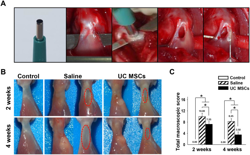
Fig 1. Procedure of surgery, macroscopic images, and quantification of macroscopic appearance of regenerated tendons at two and four weeks.
(A): Surgical procedure of FTD and intratendinous injection of saline or UC MSCs. (B): Macroscopic appearance of the tendon (Left side image: Supraspinatus tendon immediately after harvest; Right side image: the tendon removing the loose connective tissue surrounding the injury site to observe the original defect shape). (C): The total macroscopic score. Bar charts present mean ± standard deviation; Statistically significant at p < .05. Abbreviations: UC MSCs, umbilical cord derived mesenchymal stem cells.

