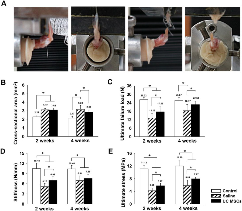
Fig 5. Biomechanical test procedure and quantification of biomechanical properties of regenerated tendons at two and four weeks.
(A): an extracted specimen and procedure of biomechanical testing. (B): Cross-sectional area of the supraspinatus tendon at defect site. (C): ultimate failure load. (D): stiffness. (E): ultimate stress. Bar charts present mean ± standard deviation; statistically significant at p < .05. Abbreviations: UC MSCs, umbilical cord derived mesenchymal stem cells.

