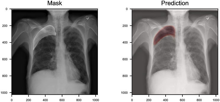
Fig 6. An example of chest radiology report.
We highlight the location of the pneumothorax lesion in the chest radiograph (left). The probabilities of segmentation output by CheXLocNet are present in varying shades of red (right). CheXLocNet correctly detected the pneumothorax and masked the lesion area roughly.

