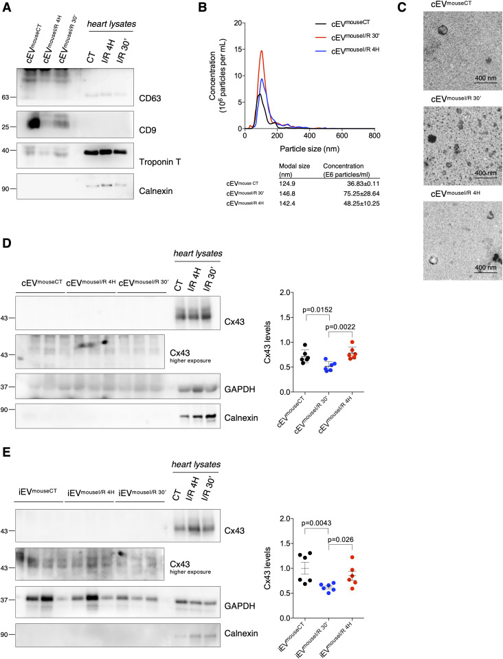
Figure 1. Ischemia decreases secretion of Cx43 into circulating extracellular vesicles (EVs) in mice subjected to myocardial I/R injury.
Left coronary artery ligation (60 min) was performed in mice, followed by reperfusion during 30 min (I/R 30′) or 4 h (I/R 4H). Sham-operated animals were used as controls (CT). (A) Non-reducing WB of circulating EVs (30 μg total protein/lane) from sham (cEVmouseCT), I/R 4H (cEVmouseI/R 4H), and I/R 30′ (cEVmouseI/R 30′). CD63 and CD9 were used as positive EV markers, Calnexin as a negative marker and Troponin T as a cardiomyocyte marker. Heart lysates were used as control. (B) Nanoparticle tracking analysis of mouse circulating EVs. (C) Representative transmission electron microscopy of mouse circulating EVs. (D) WB analysis of Cx43 in circulating EVs (30 μg total protein/lane, n = 6). GAPDH was used as a pan-EV marker. (E) WB analysis of Cx43 in intracardiac EVs (5 μg total protein/lane) from sham (iEVmouseCT), I/R 4H (iEVmouseI/R 4H), and I/R 30′ (iEVmouseI/R 30′; n = 6).
Source data are available for this figure.

