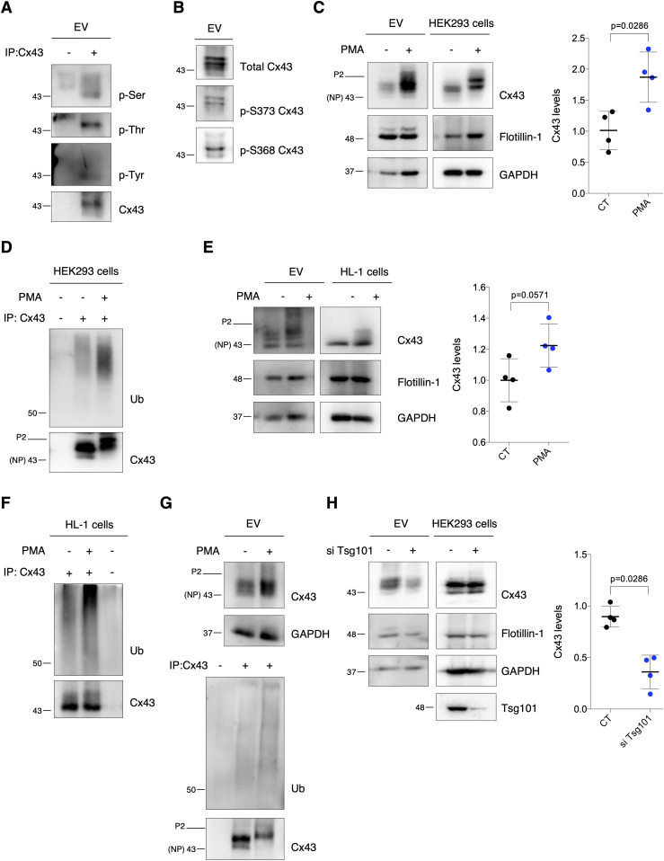
Figure 3. Extracellular vesicle (EV)-Cx43 is phosphorylated and ubiquitinated.
(A) Cx43 was immunoprecipitated (IP) from HEK293Cx43+-derived EVs. Cx43 phosphorylation was evaluated with phospho-Ser, phospho-Thr, and phospho-Tyr antibodies. (B) Phosphorylation of Cx43-S373 and Cx43-S368 was evaluated in HEK293Cx43+-derived EVs. (C) HEK293Cx43+ cells were treated with PMA or vehicle for 30 min in EV-depleted medium. EV-Cx43 levels were evaluated by WB (n = 4). (D) Cx43 ubiquitination evaluated after IP of Cx43 from HEK293Cx43+ cells. (E) HL-1 cells were treated with PMA or vehicle control for 30 min in EV-depleted medium. EV-Cx43 levels were assessed by WB (n = 4). (F) Cx43 ubiquitination evaluated after IP of Cx43 from HL-1 cells. (G) Cx43 ubiquitination evaluated after IP of Cx43 in HEK293Cx43+-derived EVs. (H) WB analysis of EV-Cx43 derived from HEK293Cx43+ cells knockdown for Tsg101 (siTsg101), incubated in EV-depleted medium for 8 h (n = 4). NP, non-phosphorylated Cx43; P2, phosphorylated Cx43.
Source data are available for this figure.

