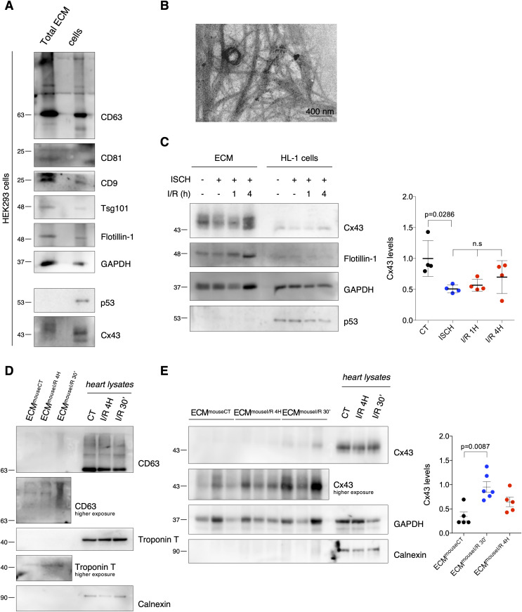
Figure 5. I/R impacts retention of extracellular vesicle (EV)-Cx43 in the ECM.
(A) Non-reducing WB of HEK293Cx43+-derived ECM (10 μg total protein/lane). CD63, CD81, CD9, Tsg101, Flotillin-1, and GAPDH was used as EV markers. p53 was used to determine cellular contamination. (B) Representative transmission electron microscopy of HEK293Cx43+-derived ECM. (C) Levels of Cx43 in ECM (10 μg total protein/lane) from HL-1 cells subjected to 30 min ischemia (ISCH), 1 h or 4 h of reperfusion (I/R; n = 4). (D) Non-reducing WB of heart-derived ECM (10 μg total protein/lane) from sham (ECMmouseCT), I/R 4H (ECMmouseI/R 4H), and I/R 30′ (ECMmouseI/R 30′). (E) WB analysis of Cx43 in heart-derived ECM (10 μg total protein/lane; n = 6).
Source data are available for this figure.

