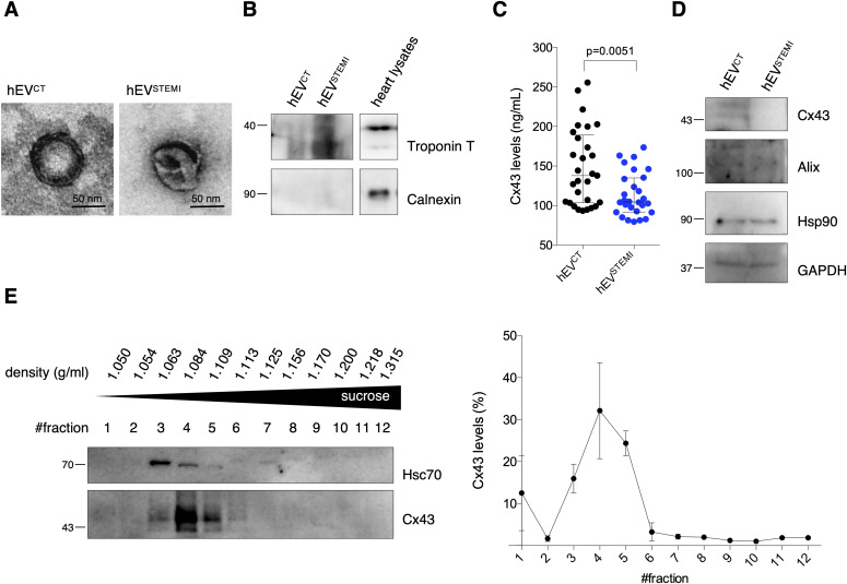
Figure 6. Cx43 levels decrease in circulating extracellular vesicles (EVs) from STEMI patients.
(A) Representative transmission electron microscopy of circulating human EVs from control (hEVCT) and STEMI patients (hEVSTEMI). (B) Representative WB of circulating EVs (30 μg total protein/lane). Heart lysates were used as control. (C) Levels of Cx43 were evaluated in hEVCT and hEVSTEMI. Individual levels, median, and interquartile range are plotted on graph (n = CT, n = 28 STEMI). (D) WB analysis of Cx43, Alix, Hsp90, and GAPDH in circulating EVs (30 μg total protein/lane). (E) Permeability of EV-Cx43 channels in circulating vesicles from human controls, assessed by sucrose-based transport-specific density shift.
Source data are available for this figure.

