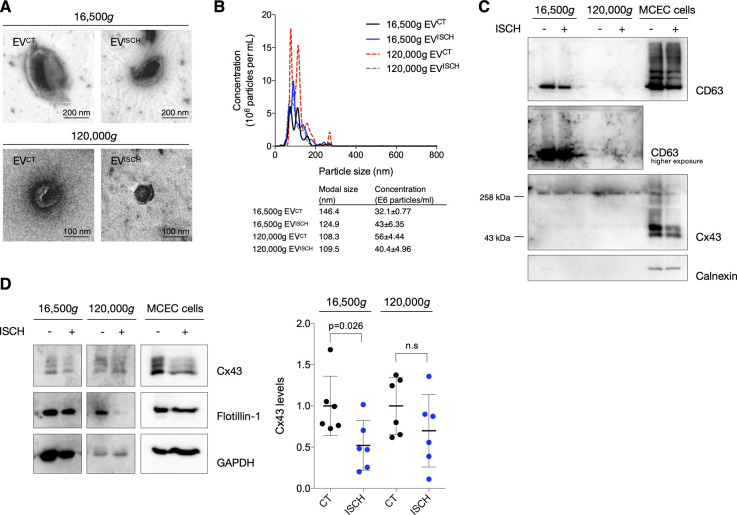
Figure S2. Ischemia does not affect Cx43 secretion into small extracellular vesicles (EVs) derived by cardiac endothelial cells.
(A) Representative transmission electron microscopy images of EVs (16,500g or 120,000g, as indicated) isolated from cardiac endothelial cells subjected or not to ischemia (ISCH). (B) Representative nanoparticle tracking analysis of concentration and size distribution of EVs derived from cardiac endothelial cells. (C) Non-reducing WB of cardiac endothelial EVs. CD63 and CD81 were used as canonical EV markers. The absence of Calnexin excludes contamination with cellular material. The presence of higher molecular bands (∼258 kD) of Cx43 denotes the presence of Cx43 hemichannels in EVs. (D) Levels of Cx43 in cardiac endothelial EVs derived from control or ischemic cells (n = 6). Analysis of the pan-EV markers Flotillin-1 and GAPDH was also performed.

