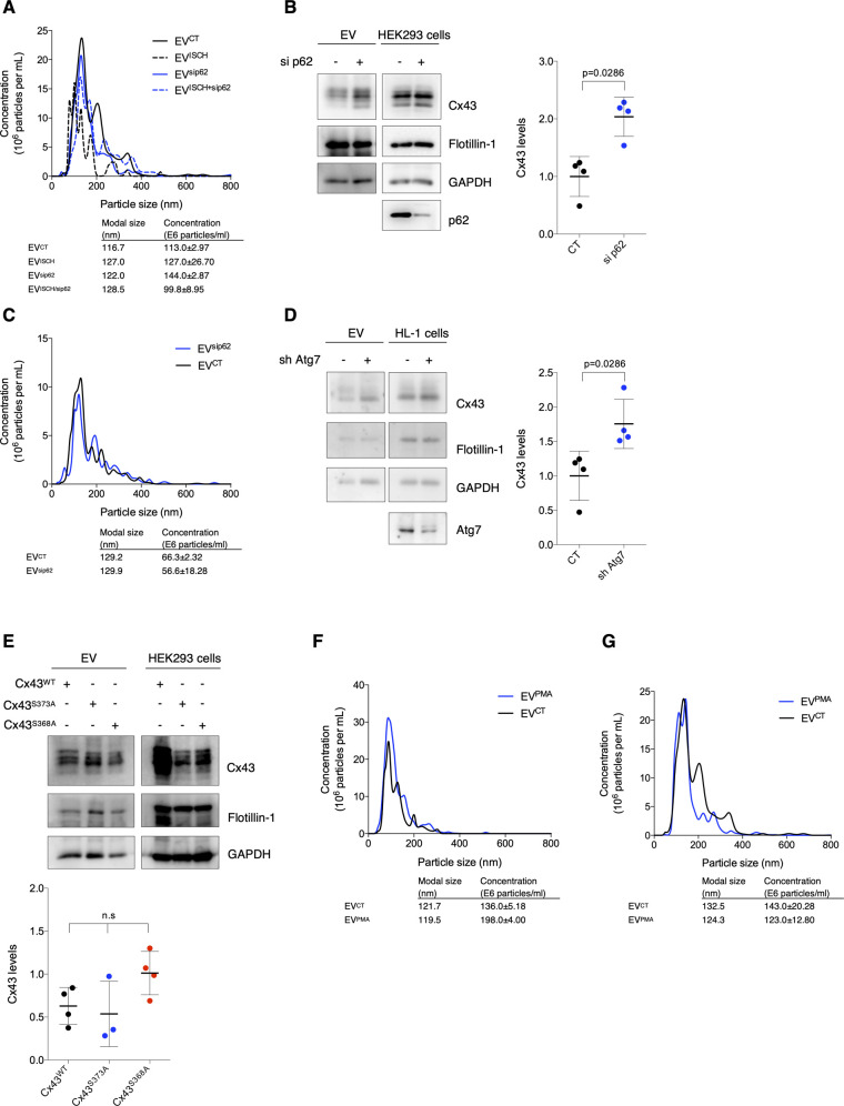
Figure S3. p62 regulates the balance between Cx43 degradation and extracellular vesicle (EV) secretion.
(A) Representative nanoparticle tracking analysis (NTA) of concentration and size distribution of EVs obtained from HL-1 cells after siRNA-mediated knockdown of p62 (sip62) and/or subjected to ischemia (ISCH). (B) knockdown of p62 (sip62) was performed in HEK293Cx43+ cells for 48 h. Cells were cultured in EV-depleted medium for 8 h, followed by EV isolation. Levels of EV-Cx43 were evaluated by WB (n = 4). Analysis of the pan-EV markers Flotillin-1 and GAPDH was also performed. (C) Representative NTA of EVs derived from HEK293Cx43+ cells depleted or not of p62 (sip62). (D) HL-1 cells were transduced with shRNA targeting Atg7 (sh Atg7), or the empty lentiviral vector (CT) for 7 d, after which cells were incubated in EV-depleted medium for 8 h. WB analysis of EV-Cx43 levels was further performed (n = 4). (E) HEK293Cx43− cells were transfected with wild type Cx43 (Cx43WT) or Cx43 mutated in the residues S373 (Cx43S373A) or S368 (Cx43S368A) for 24 h. Cells were incubated in EV-depleted medium for 8 h, followed by WB analysis of EV-Cx43 levels (n = 4). (F) Representative NTA of concentration and size distribution of EVCT and EVPMA derived from HEK293Cx43+ cells. (G) Representative NTA of concentration and size distribution of EVCT and EVPMA derived from HL-1 cells.

