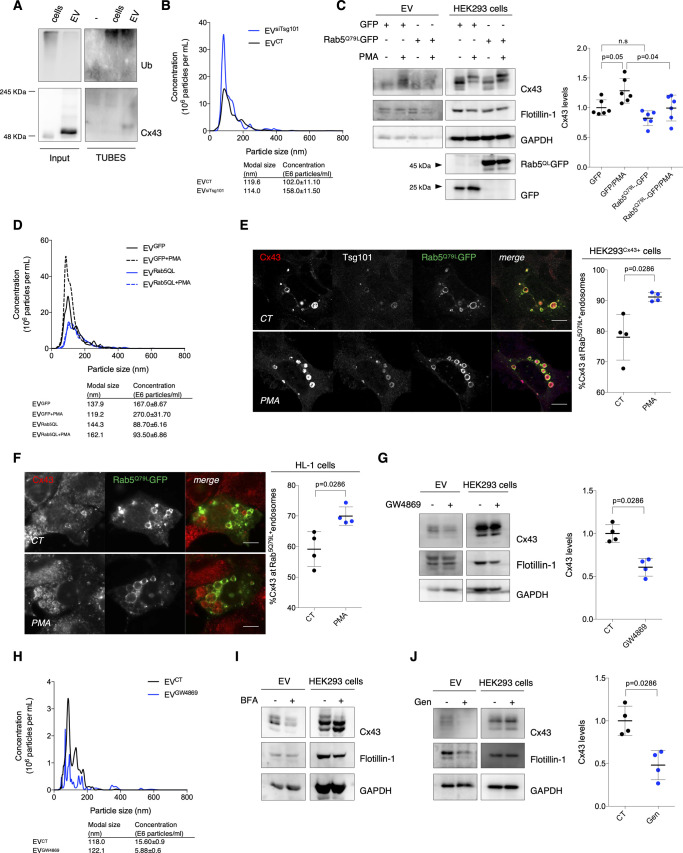
Figure S4. Cx43 found in extracellular vesicles (EVs) is of endosomal origin.
(A) Ubiquitinated substrates from lysates of EVs derived from HEK293Cx43+ cells were precipitated using TUBEs. Following elution, samples were analyzed by WB using antibodies against Cx43 and ubiquitin. Cellular extracts were used as control. (B) Representative nanoparticle tracking analysis (NTA) of concentration and size distribution of EVCT and EVsiTsg101. (C) HEK293Cx43+ cells transfected with GFP or Rab5QLGFP for 24 h, were incubated in EV-depleted medium for 8 h. Cx43 levels in EVs were evaluated by WB (n = 6). Analysis of the pan-EV markers Flotillin-1 and GAPDH was also performed. (D) Representative NTA of concentration and size distribution of EVGFP, EVGFP+PMA, EVRab5QL, and EVRab5QL+PMA. (E) HEK293Cx43+ cells transfected with Rab5QLGFP for 24 h were treated with PMA, where indicated, and immunostained for Cx43 (red) and Tsg101 (white). Nuclei were stained with DAPI. Scale bars 5 μm. Quantification of the number of Rab5QLGFP endosomes filled with Cx43 is depicted on graph (n = 4). (F) HL-1 cells transfected with Rab5QLGFP for 24 h were treated with PMA, where indicated, and immunostained for Cx43 (red). Nuclei were stained with DAPI. Scale bars 5 μm. Quantification of the number of Rab5QLGFP endosomes filled with Cx43 is depicted on graph (n = 4). (G) HEK293Cx43+ cells were treated with either GW4869 or vehicle control, for 20 h in EV-depleted medium. The levels of Cx43 were analyzed by WB (n = 4). (H) Representative NTA of concentration and size distribution of EVCT and EVGW4869. (I) HEK293Cx43+ cells were treated with either 5 μM Brefeldin A (BFA) or vehicle control, for 4 h in EV-depleted medium. The levels of Cx43 were analyzed by WB. (J) HEK293Cx43+ cells were treated with either genistein (Gen) or vehicle control, for 30 min in EV-depleted medium. The levels of Cx43 were analyzed by WB (n = 4).

