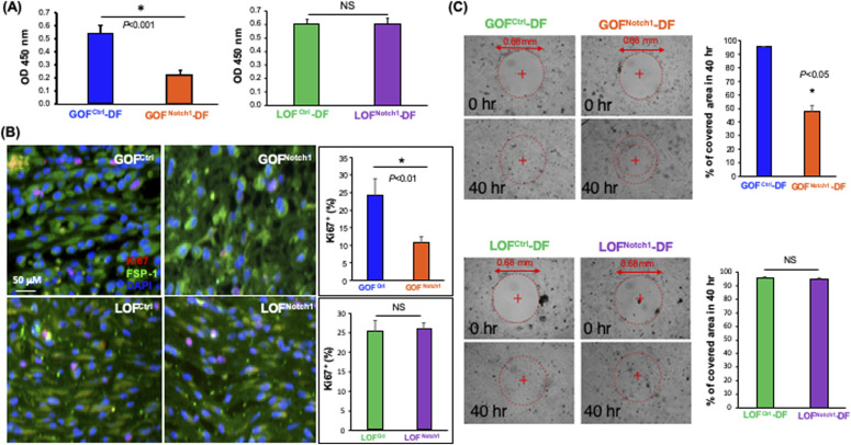
Figure 3. Activation of Notch1 pathway in fibroblasts suppresses cellular proliferation and migration.
(A) GOFNotch1-(DF) dermal fibroblast grew slower than GOFCtrl-DF, whereas growth rates of LOFNotch1-DF and LOFCtrl-DF were comparable. Data of mean ± SD are based on results of three experiments of total six wells/group (cells grew in 96-well plate) and analyzed by t test. (B) Expression of cell proliferation marker Ki67 (red) is lower in fibroblasts (FSP-1, green) at wound granulation tissue of GOFNotch1 mice than that in GOFCtrl mice, yet no obvious difference between LOFNotch1 and LOFCtrl mice. Quantitative data are calculated based on three sections/wound and mean ± SD are analyzed by t test. (C) GOFNotch1-DF migrate and proliferate were slower than those from GOFCtrl-DF, whereas migration and proliferation of LOFNotch1-DF and GOFCtrl-DF were comparable as assessed by in vitro wound healing assay. Data of mean ± SD are based on results of three experiments of total six pseudo-wounds/group and analyzed by t test.

