. 2020 Sep 21;9:e56171. doi: 10.7554/eLife.56171

Figure 7. Chronic chemogenetic activation of CamKIIα-positive forebrain excitatory neurons during the early postnatal window alters excitatory and inhibitory spontaneous currents in the hippocampi of adult male mice.

(A) Shown is a schematic of the experimental paradigm to induce chronic CNO-mediated hM3Dq DREADD activation in CamKIIα-positive forebrain excitatory neurons using bigenic CamKIIα-tTA::TetO-hM3Dq mouse pups that were fed CNO (PNCNO; 1 mg/kg) or vehicle from P2 to P14 and then left undisturbed for 3 months prior to electrophysiological analysis, in acute hippocampal slices derived from adult male mice. Whole-cell patch clamp was performed to record sEPSCs/mEPSCs and sIPSCs/mIPSCs in the somata of CA1 pyramidal neurons. R – Recording electrode. (B) Shown are representative sEPSC traces of CA1 pyramidal neurons from vehicle and PNCNO-treated mice. (C) PNCNO-treated mice showed significantly altered cumulative probability of sEPSC amplitude with a small increase at lower amplitudes (<30 pA) and a significant decline in large-amplitude events as compared to vehicle-treated controls. (D) PNCNO-treated mice showed a significant decline in the cumulative probability of sEPSC interevent intervals as compared to vehicle-treated controls (n = 8 cells for vehicle; n = 5 cells for PNCNO). (E) Shown are representative mEPSC traces of CA1 pyramidal neurons from vehicle and PNCNO-treated mice. (F) PNCNO-treated mice showed significantly enhanced cumulative probability of sEPSC amplitude as compared to vehicle-treated controls. (G) No significant change was observed in the cumulative probability of sEPSC interevent intervals in PNCNO-treated mice as compared to vehicle-treated controls (n = 5 cells for vehicle; n = 6 cells for PNCNO). (H) Shown are representative sIPSC traces of CA1 pyramidal neurons from vehicle and PNCNO-treated mice. PNCNO-treated mice showed a significant increase in the cumulative probability of sIPSC amplitude (I), along with a significant decline in the cumulative probability of sIPSC interevent intervals (J) as compared to vehicle-treated controls (n = 8 cells for vehicle; n = 7 cells for PNCNO). (K) Shown are representative mIPSC traces of CA1 pyramidal neurons from vehicle and PNCNO-treated mice. (L) PNCNO-treated mice showed a significant decline in the cumulative probability of mIPSC amplitude as compared to vehicle-treated controls (n = 6 cells for vehicle; n = 7 cells for PNCNO). (M) No significant change was observed in the cumulative probability of mIPSC interevent intervals across treatment groups. Results are expressed as cumulative probabilities. *p<0.001 as compared to PNCNO-treated group using the Kolmogorov-Smirnov two-sample comparison.

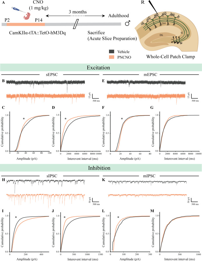
Figure 7.

Figure 7—figure supplement 1. Chronic chemogenetic activation of CamKIIα-positive forebrain excitatory neurons during the early postnatal window does not change intrinsic excitability but alters spontaneous network activity in the hippocampi of adult male mice.

Figure 7—figure supplement 1.

(A) Shown is a schematic of the experimental paradigm to induce chronic CNO-mediated hM3Dq DREADD activation in CamKIIα-positive forebrain excitatory neurons using bigenic CamKIIα-tTA::TetO-hM3Dq mouse pups that were fed CNO (PNCNO; 1 mg/kg) or vehicle from P2 to P14 and then left undisturbed for 3 months prior to electrophysiological analysis, in acute hippocampal slices derived from adult male mice. Whole-cell patch clamp was performed to record sPSCs and input-output characteristics in the somata of CA1 pyramidal neurons. R – Recording electrode. (B) Shown are representative traces of spikes generated by CA1 pyramidal neurons following a current injection of 100 pA in vehicle and PNCNO-treated adult male mice. (C) No significant change was observed in the input-output characteristics of CA1 pyramidal neurons in acute hippocampal slices derived from PNCNO-treated mice as compared to vehicle-treated controls (n = 18 cells for vehicle; n = 25 cells for PNCNO). Results are expressed as the mean ± S.E.M. (D) Shown are representative sPSC traces of CA1 pyramidal neurons from vehicle and PNCNO-treated mice. (E) PNCNO-treated mice showed significantly enhanced cumulative probability of sPSC amplitude in addition to the presence of a long-tail as compared to vehicle-treated controls. (F) PNCNO-treated mice showed a significant decline in the cumulative probability of sPSC interevent intervals as compared to vehicle-treated controls (n = 8 cells for vehicle; n = 6 cells for PNCNO). Results are expressed as cumulative probabilities. *p<0.001 as compared between vehicle and PNCNO-treated group using the Kolmogorov-Smirnov two-sample comparison.
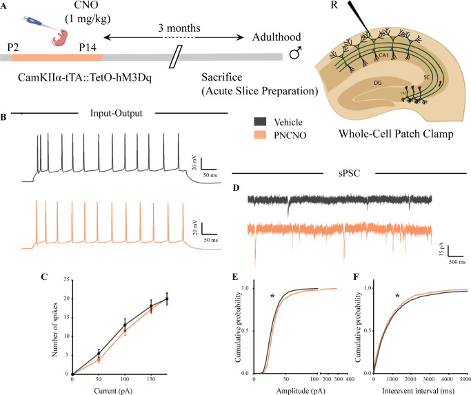
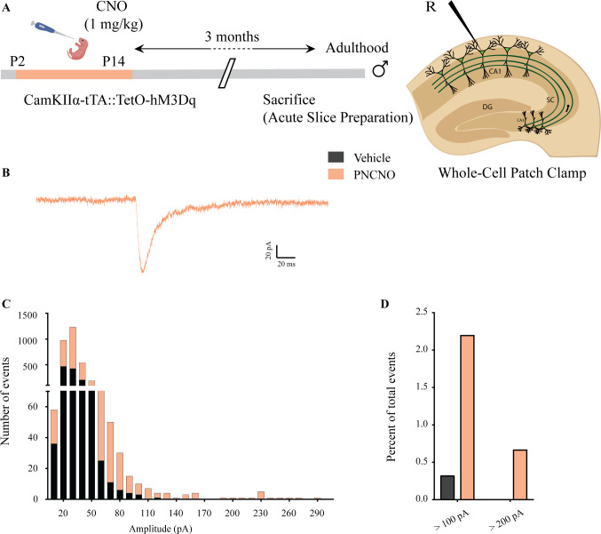
Figure 7—figure supplement 2. Effect of chronic chemogenetic activation of CamKIIα-positive forebrain excitatory neurons during the early postnatal window on the distribution of spontaneous network events in hippocampi of adult male mice.

Figure 7—figure supplement 2.

(A) Shown is a schematic of the experimental paradigm to induce chronic CNO-mediated hM3Dq DREADD activation in CamKIIα-positive forebrain excitatory neurons using bigenic CamKIIα-tTA::TetO-hM3Dq mouse pups that were fed CNO (PNCNO; 1 mg/kg) or vehicle from P2 to P14 and then left undisturbed for 3 months prior to electrophysiological analysis, in acute hippocampal slices derived from adult male mice. Whole-cell patch clamp was performed to record sPSCs in the somata of CA1 pyramidal neurons. R – Recording electrode. (B) Shown is an example trace of a large-amplitude spontaneous network event observed in a CA1 pyramidal neuron of a PNCNO-treated mouse. (C) Distribution plot for sPSC amplitudes showing a long-tail of large-amplitude events in PNCNO-treated mice. (D) Quantification of percent of total events with amplitude >100 pA and >200 pA. No event >200 pA was observed in vehicle-treated controls.