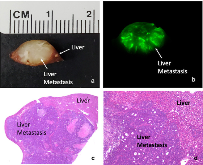
Figure 3. Ex-vivo imaging of a tumor section. (a) Bright-light imaging of tumor with surrounding liver. Tumor measures approximately 1 cm in diameter. (b) Ex-vivo fluorescence imaging of a metastasis section demonstrating penetration of the huCC49-IR800 antibody throughout the entire depth of the metastasis. (c) H&E staining of liver metastasis visualized using a 2× objective lens. (d) H&E staining of liver metastasis using a 10× objective lens.