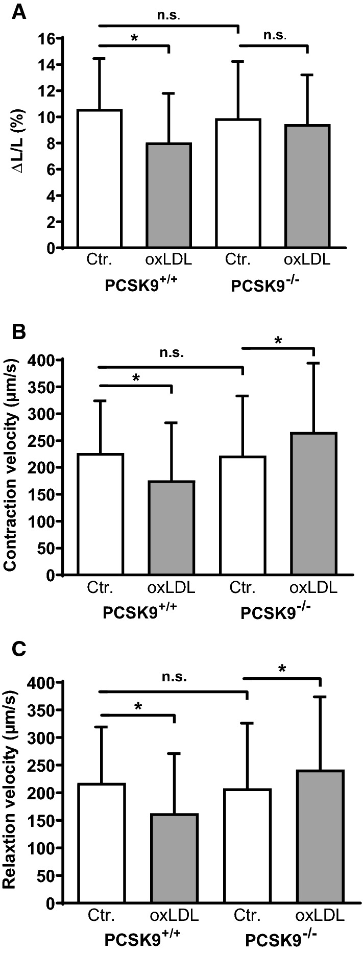
Fig. 1.

Effect of oxLDL on load-free cell shortening of isolated cardiomyocytes derived from PCSK9−/−- and PCSK9+/+-mice. Serum-free cultured adult ventricular cardiomyocytes were exposed to 5 µM oxLDL for 24 h. Load free cell shortening (cells were paced at 2 Hz) is expressed (a) as ΔL/L (%), (b) contraction velocity (µm/s) (c) and relaxation velocity (µm/s) of 222 (PCSK9+/+, Control), 126 (PCSK9+/+, oxLDL), 162 (PCSK9−/−, Control) and 134 (PCSK9−/−, oxLDL) cells (14–25 independent experiments with an intraassay variability of p > 0.05). Statistical analysis was performed by Mann–Whitney test. *p ≤ 0.05, n.s. not significant. Data are mean ± SD
