Fig. 10.

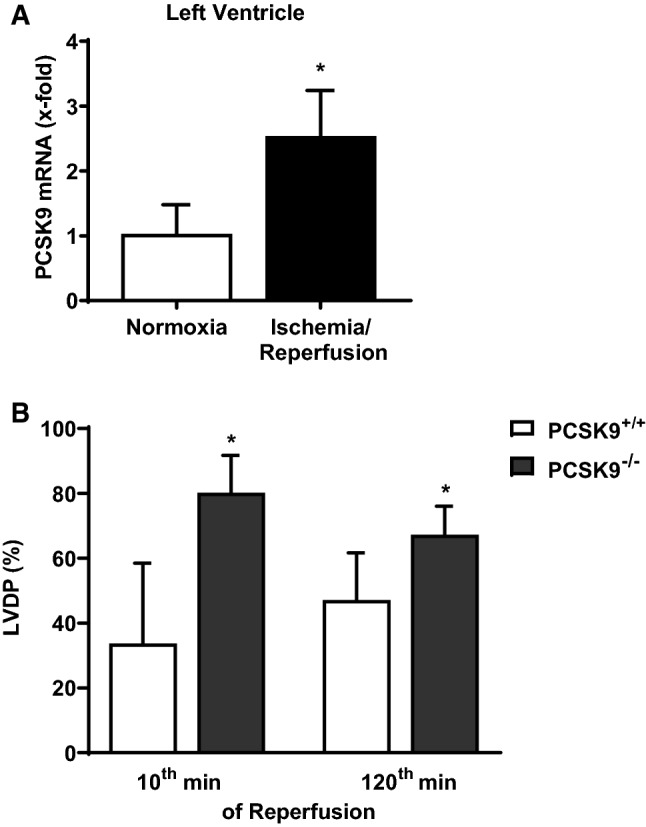
PCSK9 expression in ischemic mice hearts as well as left ventricular function of PCSK9 knockout hearts. a Hearts from C57BL6/JR mice were excised and exposed to 45 min of ischemia and 120 min reperfusion (I/R) or 165 min normoxia (Nx). PCSK9 mRNA expression of left ventricles was analyzed and normalized to the mean expression of B2M, HPRT and GAPDH. Nx: n = 10, I/R: n = 9. Statistical analysis was performed by unpaired t-test. *p ≤ 0.05. b Left ventricular developed pressure (LVDP) of the 10th minute as well as of the 120th minute of reperfusion from isolated hearts (PCSK9−/−- and PCSK9+/+-mice) was measured and normalized to the LVDP under basal conditions (given in %). n = 11–13. Statistical analysis was performed by unpaired t-test. *p ≤ 0.05. All data are mean ± SD
