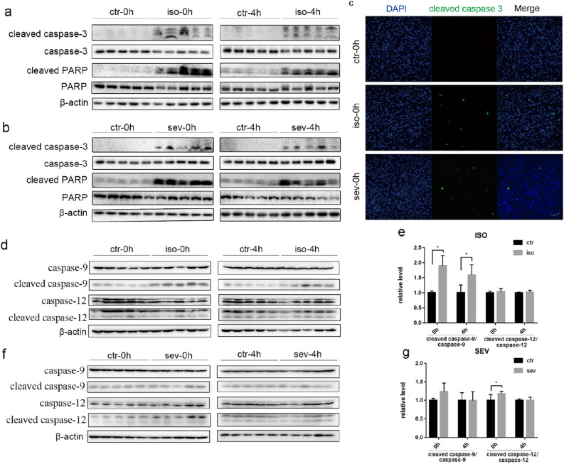
Figure 1.
Neuronal Apoptosis in Neonatal Mice following Exposure to Isoflurane or Sevoflurane. (a, b, d, f) Brain homogenates from neonatal mice sacrificed at 0 h and 4 h after exposure to anesthesia were analyzed by western blotting using antibodies against cleaved caspase-3, caspase-3, cleaved PARP, PARP, cleaved caspase-9, caspase-9, cleaved caspase-12, caspase-12 and β-actin. Full-length blots are presented in Supplementary Fig. 3. (c) Representative immunofluorescence staining of the brain cortex from three groups immediately after treatment (scale bar, 50 μm). (e, g) Densitometric quantification of western blots after normalization to β-actin levels. Values are presented as mean ± SD. *P < 0.05 vs. Control. n = 5 per group. Ctr control, iso isoflurane, sev sevoflurane.

