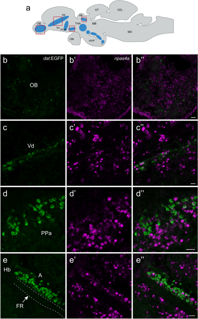
Figure 5.
Expression of npas4a gene in the brain of transgenic dat:EGFP fish followed by 30-min post Kiss1 administration. (a) Schematic sagittal view of a zebrafish brain illustrating the distribution of dopaminergic cell population (blue-shaded zones) in the brain of zebrafish. Boxes with red indicate the boundaries of the photomicrographic illustrations in below (b–e). (b–e) EGFP (green)-labelled dopaminergic neurons in the olfactory bulb (OB, b); dorsal nucleus of ventral telencephalic area (Vd, c); anterior part of the parvocellular preoptic nucleus (PPa, d) and in the anterior thalamic nucleus (A, e) above the fasciculus retroflexus (FR). Although cells expressing npas4a mRNA (magenta) were seen in several brain regions (b'–e'), there was no expression of npas4a in dopaminergic cells (b"−e"). Scale bars 20 µm.

