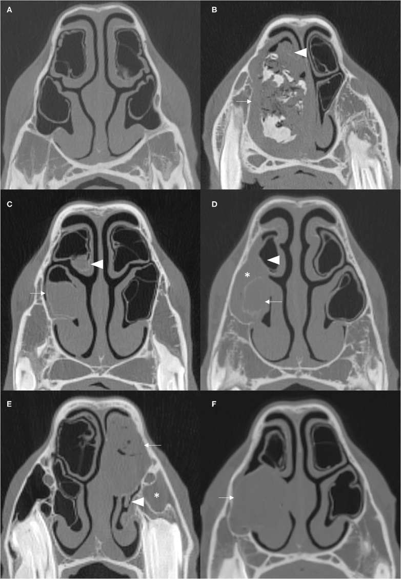
Figure 1.
Transverse CT images of normal NCBs and of various types of NCB empyema. (A) Normal NCBs. (B) Mixed hyper- and hypoattenuation within a distended VCB, representing mineralisation and gas, respectively, within soft tissue attenuating material (arrow). There is compression and severe damage of the ipsilateral DCB (arrowhead) (mineralised nasal conchae found on histology—disorder of 10 years clinical duration). (C) Soft tissue/fluid attenuating material fills most of VCB (arrow) and the ventral aspect of DCB (arrowhead). (D) Soft tissue/fluid attenuating material fills the entire VCB (arrow), which has a thickening of the bony concha and has surrounding soft tissue/fluid attenuating material flowing from the ipsilateral sinuses (asterisk) and mild damage to the ipsilateral DCB (arrowhead). (E) The DCB is partially filled with material of mixed soft tissue and gas attenuation, reflecting inspissated purulent exudate (arrow). There is moderate damage of the ipsilateral VCB (arrowhead) and ipsilateral sinus empyema (asterisk). (F) The VCB is distended with homogenous soft tissue/fluid attenuating material (arrow). All CT images were reconstructed using using a bone filter (Window Level 800 HU, Window Width 2,800 HU). The right side of the patient is on the left side of the image. The transverse images (A–C) are at the level of the Triadan 08 maxillary cheek teeth, (D,F) at the level of the Triadan 07s and (E) is level with the distal (caudal) aspect of Triadan 08s.

