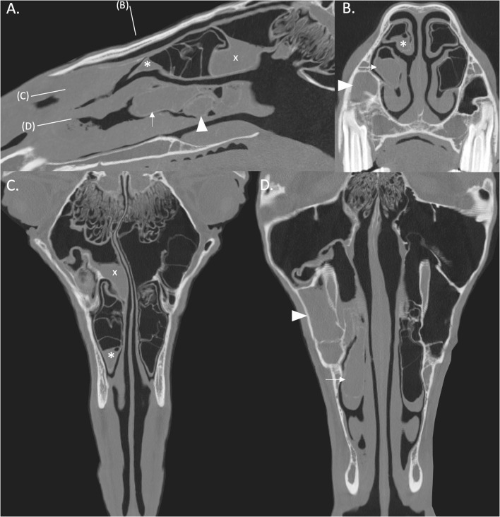
Figure 2.
(A) Right parasagittal CT reconstruction with lines representing the locations of images (B–D). (B) Transverse CT image. (C,D) Dorsal CT reconstruction at the level of the DCB and VCB, respectively, the rostral aspect is toward the bottom of the image. There is empyema of the VCB (arrow) with ipsilateral sinusitis of the rostral (arrowhead) and caudal (x) paranasal sinus compartments. There is thickening of the mucosa of the rostral aspect of the right DCB (asterisk). All CT images are displayed using a bone filter (Window Level 800 HU, Window Width 2,800 HU). The right side of the patient is on the left side of the image.

