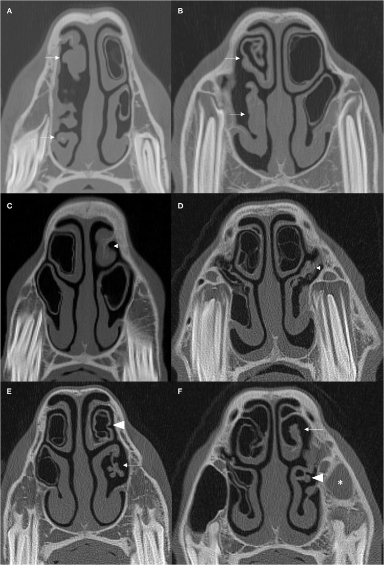
Figure 3.
(A,B) Transverse CT images of two cases with moderate to severe damage of ipsilateral DCBs and VCBs (arrows) with distortion of the adjacent nasal concha. (C) The left DCB is not present (arrow) and there is contraction and thickening of the remaining adjacent nasal concha. (D) There is loss of the left VCB (arrow) with flattening and irregular thickening of the surrounding ventral nasal concha. (E) There is loss of the left VCB (arrow) with distortion and atrophy of the lateral aspect of the surrounding ventral concha. The walls of the ipsilateral DCB is hyper-attenuated and has a scalloped appearance (arrowhead). (F) There is loss of the DCB (arrow) and distortion and thickening of the adjacent concha and loss of identifiable structure in the VCB (arrowhead). There is soft tissue/fluid attenuation filling the left rostral maxillary sinus consistent with ipsilateral sinusitis (asterisk). All CT images are displayed using a bone filter (Window Level 800 HU, Window Width 2,800 HU). The right side of the patient is on the left side of the image. Transverse images (A,E) are at the level of the Triadan 07 maxillary cheek teeth, (B–D) at the level of the Triadan 08s and (F) is level with the distal (caudal) aspect of the Triadan 08s.

