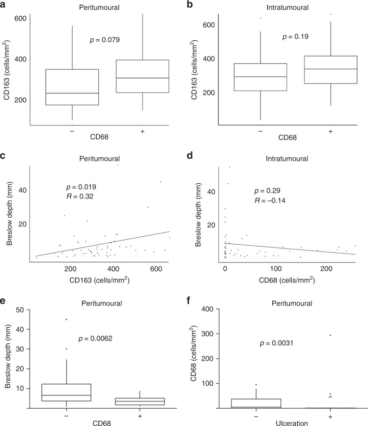
Fig. 1. Differential CD68+ and CD163+ macrophage recruitment to primary melanoma lesions.
The number of CD68+ and CD163+ cells was determined by immunohistochemistry in primary melanoma tumours. The number of positive cells was counted in the peritumoural and intratumoural regions. CD68+ and CD163+ cells were shown to be differentially recruited (a, b). Intratumoural CD163+ macrophage infiltration was positively correlated to Breslow depth (c), while intratumoural CD68+ macrophage infiltration showed no significant correlation (d). Tumours with no peritumoural CD68+ macrophages had a greater Breslow depth (e), and decreased numbers of peritumoural CD68+ macrophages were seen in ulcerated tumours (f).

