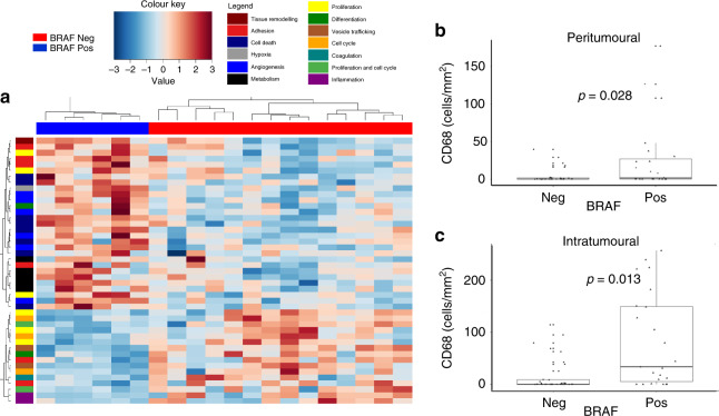
Fig. 6. The effect of BRAF status on CD68+ macrophage density and gene expression.
The number of CD68+ macrophages was determined by immunohistochemistry in primary melanoma tumours. a Intratumoural gene expression of the same FFPE blocks was measured using next-generation sequencing technology. The BRAF status of samples is shown along the upper margin with BRAF mutation-positive samples marked blue and BRAF mutation-negative samples marked red. Using Deseq2, genes differentially expressed based on BRAF status were identified. Gene-associated signalling pathways are colour coded on the left-hand margin. Using Wilcoxon rank-sum tests, it was shown that there were a higher number of peritumoural and intratumoural CD68+ macrophages in BRAF+ tumours (b, c).

