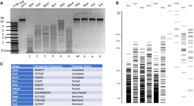
FIGURE 2.
Type II restriction of phage T4gt DNA. Phage T4gt gDNA was digested by 10 REases and analyzed by agarose gel electrophoresis (A). 2 log, DNA size marker in 100 bp to 10 kb (NEB). The predicted digestion patterns by NEBcutter are shown in (B). The target recognition sequence and digestion results are summarized in (C). C = complete digestion, P = partial digestion, VP = very partial digestion (only a few weak bands), X = resistant to restriction.

