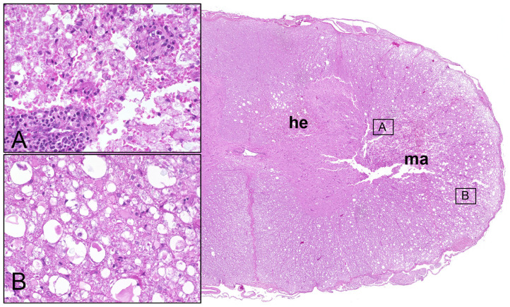
Figure 1.
Male Dachshund with type I intervertebral disc herniation (acute extrusion). Overview (right side) of HE stained spinal cord transversal section with hemorrhage (he) accentuated within the gray matter and white matter malacia (ma). Inset upper left (A): moderate perivascular cuffing of mononuclear leukocytes and focal disintegration of neuroparenchyma (necrosis, malacia). Inset lower left (B): moderate to severe white matter vacuolation within the ventrolateral funiculus, characterized by multiple dilated myelin sheaths that contain hypereosinophilic swollen axons (spheroids). 20x magnification in insets.

