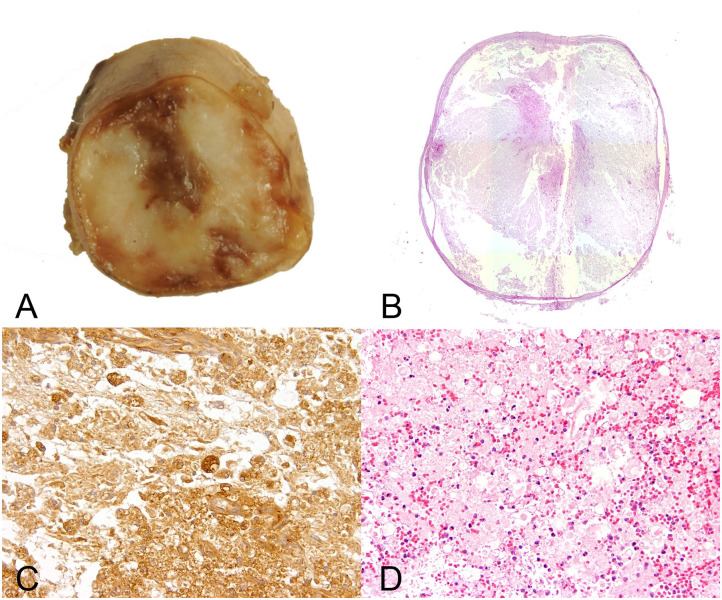
Figure 4.
Male 6 years old Yorkshire Terrier with progressive myelomalacia (PMM) following acute intervertebral disc extrusion. In PMM the shown lesions are not restricted to the initial site of spinal cord injury but extend several centimeters into cranial and caudal direction (ascending and descending malacia). (A) Gross picture of a transversal section of the formalin fixed spinal cord with complete disintegration of spinal cord neuroparenchyma and hemorrhage. (B) The HE stained overview of the transversal section shows polio- and leukomyelomalacia with complete loss of cellular details and loss of distinction between white and gray matter. (C) Multiple foamy microglia/macrophages labeled by the lectin of Bandeiraea simplicifolia 1 have infiltrated the lesion and remove cellular debris. 40x magnification. (D) There is severe extravasation of erythrocytes within the white and gray matter (hemorrhage), associated with infiltration of viable and degenerate neutrophils adjacent to areas of white matter damage with spheroids and myelin vacuolation. 10x magnification.

