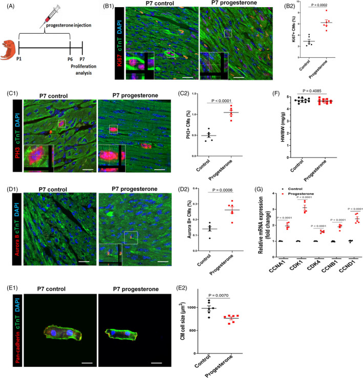
Figure 2.

Progesterone supplementation promotes postnatal CM proliferation in vivo. A, Overview of the experimental set‐up of the daily intraperitoneal injection of progesterone (8 mg/kg) or control vehicle (corn oil) from P1 to P6. Hearts were harvested at P7. B‐D, CM proliferation was evaluated by Ki67, PH3 and Aurora B immunostaining. Representative images with z‐stacking are shown in B1, C1 and D1, and quantification of percentages of Ki67+, PH3+ or Aurora B+ CMs is shown in B2, C2 and D2, respectively (n = 6). Scale bars are 20 µm. E, Representative images (E1) and quantification (E2) of the cell size of CMs (labelled by cTnT and pan‐cadherin to show intact cardiomyocytes) isolated from P7 heart. Eighty to one hundred cells were randomly selected per heart (n = 6). Scale bars are 20 µm. F, Heart weight and body weight (HW/BW) ratio (n = 10‐11). G, The mRNA expression of cell cycle activators in mouse hearts was analysed by real‐time PCR (n = 5). CM, cardiomyocyte
